Explore
 Validate
 Learn
Learn Western blot
 Immunoprecipitation
Antibody data
- Antibody Data
- Antigen structure
- References [0]
- Comments [0]
- Validations
- Western blot [1]
- Immunocytochemistry [3]
Submit
Validation data
Reference
Comment
Report error
- Product number
- SM3108P - Provider product page

- Provider
- Acris Antibodies GmbH
- Proper citation
- Acris Antibodies GmbH Cat#SM3108P, RRID:AB_1001433
- Product name
- anti FYN
- Antibody type
- Monoclonal
- Antigen
- Bacterially expressed recombinant fragment of human Fyn (aa 7-176)
- Reactivity
- Human, Mouse
- Host
- Mouse
- Isotype
- IgG
- Antibody clone number
- FYN-01
- Vial size
- 0.1 mg
- Concentration
- 1.0 mg/ml
No comments: Submit comment
Supportive validation
- Submitted by
- Acris Antibodies GmbH (provider)
- Main image
- Experimental details
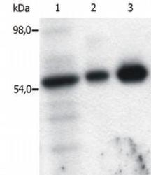
- Figure 4. Immunoprecipitation of Fyn from the lysate of T cells isolated from fresh buffy coats. Western blot was immunostained with anti-Fyn (FYN-01). Lane 1: original lysate of T cells. Lane 2-3: Immunoprecipitated material eluted from affinity sorbent (FYN-01 coupled to Sepharose beads). Lanes differ in amount of T cell lysate loaded on the immunosorbent.

Supportive validation
- Submitted by
- Acris Antibodies GmbH (provider)
- Main image
- Experimental details
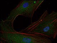
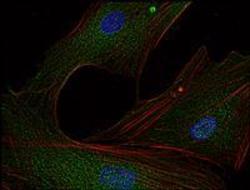
- Figure 1. Immunofluorescence staining of Fyn in murine transformed fibroblasts using anti-Fyn (FYN-01; red). Actin cytoskeleton was decorated by phalloidin (green) and cell nuclei stained with DAPI (blue).
- Submitted by
- Acris Antibodies GmbH (provider)
- Main image
- Experimental details
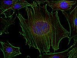
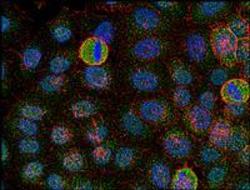
- Figure 2. Immunofluorescence staining of Fyn in human HeLa cell line using anti-Fyn (FYN-01; green). Actin cytoskeleton was decorated by phalloidin (red) and cell nuclei stained with DAPI (blue).
- Submitted by
- Acris Antibodies GmbH (provider)
- Main image
- Experimental details
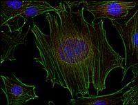
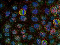
- Figure 3. Immunofluorescence staining of Fyn in human primary fibroblasts using anti-Fyn (FYN-01; green). Actin cytoskeleton was decorated by phalloidin (red) and cell nuclei stained with DAPI (blue).