Explore
 Validate
 Learn
Learn Western blot
Antibody data
- Antibody Data
- Antigen structure
- References [4]
- Comments [0]
- Validations
- Western blot [2]
- Immunocytochemistry [1]
- Immunohistochemistry [1]
Submit
Validation data
Reference
Comment
Report error
- Product number
- AF3197 - Provider product page

- Provider
- R&D Systems
- Product name
- Human/Mouse/Rat AMPK alpha 1 Antibody
- Antibody type
- Polyclonal
- Description
- Antigen Affinity-purified. Detects human, mouse, and rat AMPK alpha 1 in Western blots. The antibody does not cross-react with recombinant human AMPK alpha 2.
- Reactivity
- Human, Mouse, Rat
- Host
- Goat
- Conjugate
- Unconjugated
- Antigen sequence
Q13131- Isotype
- IgG
- Vial size
- 100 ug
- Concentration
- LYOPH
- Storage
- Use a manual defrost freezer and avoid repeated freeze-thaw cycles. 12 months from date of receipt, -20 to -70 °C as supplied. 1 month, 2 to 8 °C under sterile conditions after reconstitution. 6 months, -20 to -70 °C under sterile conditions after reconstitution.
Submitted references Disrupted structure and aberrant function of CHIP mediates the loss of motor and cognitive function in preclinical models of SCAR16.
Modulating the therapeutic response of tumours to dietary serine and glycine starvation.
AMP-activated protein kinase modulates tau phosphorylation and tau pathology in vivo.
Regulation of AMP-activated protein kinase signaling by AFF4 protein, member of AF4 (ALL1-fused gene from chromosome 4) family of transcription factors, in hypothalamic neurons.
Shi CH, Rubel C, Soss SE, Sanchez-Hodge R, Zhang S, Madrigal SC, Ravi S, McDonough H, Page RC, Chazin WJ, Patterson C, Mao CY, Willis MS, Luo HY, Li YS, Stevens DA, Tang MB, Du P, Wang YH, Hu ZW, Xu YM, Schisler JC
PLoS genetics 2018 Sep;14(9):e1007664
PLoS genetics 2018 Sep;14(9):e1007664
Modulating the therapeutic response of tumours to dietary serine and glycine starvation.
Maddocks ODK, Athineos D, Cheung EC, Lee P, Zhang T, van den Broek NJF, Mackay GM, Labuschagne CF, Gay D, Kruiswijk F, Blagih J, Vincent DF, Campbell KJ, Ceteci F, Sansom OJ, Blyth K, Vousden KH
Nature 2017 Apr 19;544(7650):372-376
Nature 2017 Apr 19;544(7650):372-376
AMP-activated protein kinase modulates tau phosphorylation and tau pathology in vivo.
Domise M, Didier S, Marinangeli C, Zhao H, Chandakkar P, Buée L, Viollet B, Davies P, Marambaud P, Vingtdeux V
Scientific reports 2016 May 27;6:26758
Scientific reports 2016 May 27;6:26758
Regulation of AMP-activated protein kinase signaling by AFF4 protein, member of AF4 (ALL1-fused gene from chromosome 4) family of transcription factors, in hypothalamic neurons.
Komori T, Doi A, Nosaka T, Furuta H, Akamizu T, Kitamura T, Senba E, Morikawa Y
The Journal of biological chemistry 2012 Jun 8;287(24):19985-96
The Journal of biological chemistry 2012 Jun 8;287(24):19985-96
No comments: Submit comment
Supportive validation
- Submitted by
- R&D Systems (provider)
- Main image

- Experimental details
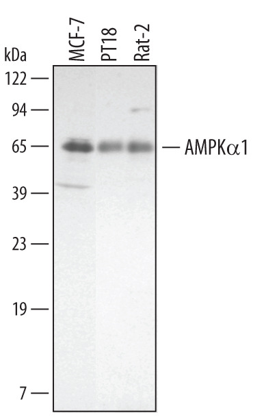
- Detection of Human/Mouse/Rat AMPK alpha 1 by Western Blot. Western blot shows lysates of MCF-7 human breast cancer cell line, PT18 mouse mast/basophil cell line, and Rat-2 rat embryonic fibroblast cell line. PVDF membrane was probed with 0.5 µg/mL of Goat Anti-Human/Mouse/Rat AMPK alpha 1 Antigen Affinity-purified Polyclonal Antibody (Catalog # AF3197) followed by HRP-conjugated Anti-Goat IgG Secondary Antibody (Catalog # HAF109). A specific band was detected for AMPK alpha 1 at approximately 63 kDa (as indicated). This experiment was conducted under reducing conditions and using Immunoblot Buffer Group 1.
- Submitted by
- R&D Systems (provider)
- Main image

- Experimental details
- Detection of Human. Mouse, and Rat AMPK alpha 1 by Western Blot. Western blot shows lysates of HeLa human cervical epithelial carcinoma cell line, C2C12 mouse myoblast cell line, and Rat-2 rat embryonic fibroblast cell line. PVDF membrane was probed with 2 µg/mL of Goat Anti-Human/Mouse/Rat AMPK alpha 1 Antigen Affinity-purified Polyclonal Antibody (Catalog # AF3197) followed by HRP-conjugated Anti-Goat IgG Secondary Antibody (Catalog # HAF017). A specific band was detected for AMPK alpha 1 at approximately 70 kDa (as indicated). This experiment was conducted under reducing conditions and using Immunoblot Buffer Group 1.

Supportive validation
- Submitted by
- R&D Systems (provider)
- Main image

- Experimental details
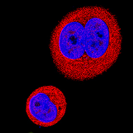
- AMPK alpha 1 in MCF-7 Human Cell Line. AMPK alpha 1 was detected in immersion fixed MCF-7 human breast cancer cell line using Goat Anti-Human/Mouse/Rat AMPK alpha 1 Antigen Affinity-purified Polyclonal Antibody (Catalog # AF3197) at 5 µg/mL for 3 hours at room temperature. Cells were stained using the NorthernLights™ 557-conjugated Anti-Goat IgG Secondary Antibody (red; Catalog # NL001) and counterstained with DAPI (blue). Specific staining was localized to nuclei and cytoplasm. View our protocol for Fluorescent ICC Staining of Cells on Coverslips.
Supportive validation
- Submitted by
- R&D Systems (provider)
- Main image

- Experimental details
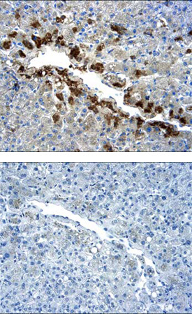
- AMPK alpha 1 in Human Liver. AMPK alpha 1 was detected in immersion fixed paraffin-embedded sections of human liver using 5 µg/mL Goat Anti-Human/Mouse/Rat AMPK alpha 1 Antigen Affinity-purified Polyclonal Antibody (Catalog # AF3197) overnight at 4 °C. Tissue was stained with the Anti-Goat HRP-DAB Cell & Tissue Staining Kit (brown; Catalog # CTS008) and counterstained with hematoxylin (blue). Lower panel shows secondary antibody only control experiment. View our protocol for Chromogenic IHC Staining of Paraffin-embedded Tissue Sections.