Explore
 Validate
 Learn
Learn Western blot
Antibody data
- Antibody Data
- Antigen structure
- References [0]
- Comments [0]
- Validations
- Western blot [1]
- Immunocytochemistry [1]
- Immunohistochemistry [1]
- Flow cytometry [1]
Submit
Validation data
Reference
Comment
Report error
- Product number
- TA300537 - Provider product page

- Provider
- OriGene
- Proper citation
- OriGene Cat#TA300537, RRID:AB_2105619
- Product name
- Rabbit Monoclonal Antibody against FRAP1 (Clone Y391)
- Antibody type
- Monoclonal
- Description
- Rabbit Monoclonal Antibody against FRAP1 (Clone Y391)
- Host
- Rabbit
- Conjugate
- Unconjugated
- Epitope
- MTOR
- Isotype
- IgG
- Antibody clone number
- Y391
- Vial size
- 100 µl
- Concentration
- NULL
No comments: Submit comment
Supportive validation
- Submitted by
- OriGene (provider)
- Main image

- Experimental details
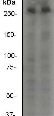
- Western blot - mTOR antibody [Y391]; All lanes : Anti-mTOR antibody [Y391] - ChIP Grade at 1/2000 dilution.Lane 1 : Hela cell lysate.Lane 2 : MCF-7.Predicted band size : 289 kDa.Observed band size : >250 kDa .
- Validation comment
- WB
Supportive validation
- Submitted by
- OriGene (provider)
- Main image
- Experimental details
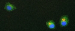
- ICC/IF image of TA300537 stained MCF7 cells. The cells were incubated with the antibody overnight at 4.. The secondary antibody (green) was Alexa Fluor 488 goat anti-rabbit IgG (H+L) used at 1:1000 for 1h. Alexa Fluor 594 WGA was used to label plasma membranes (red) at 1:200 for 1h. DAPI was used to stain the cell nuclei (blue) at a concentration of 1.43uM.
- Validation comment
- IF

Supportive validation
- Submitted by
- OriGene (provider)
- Main image
- Experimental details
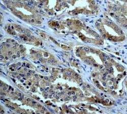
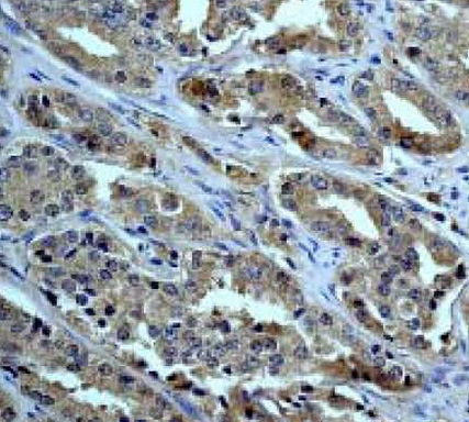
- Immunohistochemistry (Paraffin-embedded sections) - mTOR antibody [Y391]; Immunohistochemical analysis of mTOR expression in paraffin-embedded human prostate carcinoma, using 1/250 TA300537.
- Validation comment
- IHC
Supportive validation
- Submitted by
- OriGene (provider)
- Main image

- Experimental details
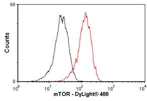
- Flow Cytometry-mTOR antibody- ChIP Grade(TA300537); Overlay histogram showing HeLa cells stained with TA300537 (red line). The secondary antibody used was DyLight 488 goat anti-rabbit IgG (H+L) at 1:500. Isotype control antibody (black line) was rabbit monoclonal IgG (1ug/1x10^6 cells) used under the same conditions. This antibody gave a significantly decreased signal in HeLa cells fixed with 4% paraformaldehyde (10 min)/permeabilized with 0.1% PBS-Tween used under the same conditions.
- Validation comment
- FC