 Explore
 Validate
 Learn
LearnPA5-29026
antibody from Invitrogen Antibodies
Targeting: HDAC3
HD3, KDAC3, RPD3, RPD3-2
 Western blot Immunocytochemistry
 Immunoprecipitation Immunohistochemistry Chromatin Immunoprecipitation Other assay
Antibody data
- Antibody Data
- Antigen structure
- References [1]
- Comments [0]
- Validations
- Western blot [8]
- Immunocytochemistry [2]
- Immunohistochemistry [1]
- Chromatin Immunoprecipitation [1]
- Other assay [1]
Submit
Validation data
Reference
Comment
Report error
- Product number
- PA5-29026 - Provider product page

- Provider
- Invitrogen Antibodies
- Product name
- HDAC3 Polyclonal Antibody
- Antibody type
- Polyclonal
- Antigen
- Synthetic peptide
- Description
- Recommended positive controls: 293T, A431, H1299, HeLaS3, HepG2, Molt-4, Raji, U87-MG, SK-N-SH, IMR32, SK-N-AS, Neuro2A, C8D30, NIH-3T3, Raw264.7, C2C12, PC-12, Rat2, FaDu. Predicted reactivity: Mouse (100%), Rat (100%), Pig (100%), Chicken (100%), Rhesus Monkey (100%), Bovine (100%). Store product as a concentrated solution. Centrifuge briefly prior to opening the vial.
- Reactivity
- Human, Mouse, Rat
- Host
- Rabbit
- Isotype
- IgG
- Vial size
- 100 µL
- Concentration
- 0.66 mg/mL
- Storage
- Store at 4°C short term. For long term storage, store at -20°C, avoiding freeze/thaw cycles.
Submitted references Transcriptomic and genomic studies classify NKL54 as a histone deacetylase inhibitor with indirect influence on MEF2-dependent transcription.
Minisini M, Di Giorgio E, Kerschbamer E, Dalla E, Faggiani M, Franforte E, Meyer-Almes FJ, Ragno R, Antonini L, Mai A, Fiorentino F, Rotili D, Chinellato M, Perin S, Cendron L, Weichenberger CX, Angelini A, Brancolini C
Nucleic acids research 2022 Mar 21;50(5):2566-2586
Nucleic acids research 2022 Mar 21;50(5):2566-2586
No comments: Submit comment
Supportive validation
- Submitted by
- Invitrogen Antibodies (provider)
- Main image

- Experimental details
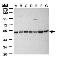
- Western Blot using HDAC3 Polyclonal Antibody (Product # PA5-29026). Sample (30 µg whole cell lysate). A: 293T. B: A431. C: H1299. D: HeLa S3. E: HepG2. F: MOLT4. G: Raji. 10% SDS PAGE. HDAC3 Polyclonal Antibody (Product # PA5-29026) diluted at 1:1,000. The HRP-conjugated anti-rabbit IgG antibody was used to detect the primary antibody.

- Submitted by
- Invitrogen Antibodies (provider)
- Main image
- Experimental details
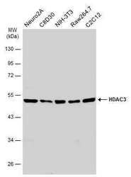
- Western Blot using HDAC3 Polyclonal Antibody (Product # PA5-29026). Various whole cell extracts (30 µg) were separated by 10% SDS-PAGE, and the membrane was blotted with HDAC3 Polyclonal Antibody (Product # PA5-29026) diluted at 1:1,000. The HRP-conjugated anti-rabbit IgG antibody was used to detect the primary antibody.

- Submitted by
- Invitrogen Antibodies (provider)
- Main image
- Experimental details
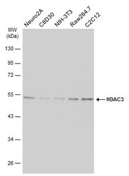
- Western Blot analysis of HDAC3 was performed by separating 30 µg of various whole cell extracts by 10% SDS-PAGE. Proteins were transferred to a membrane and probed with a HDAC3 Polyclonal Antibody (Product # PA5-29026) at a dilution of 1:1000 and a HRP-conjugated anti-rabbit IgG secondary antibody.

- Submitted by
- Invitrogen Antibodies (provider)
- Main image
- Experimental details
- Western Blot using HDAC3 Polyclonal Antibody (Product # PA5-29026). Sample (30 µg whole cell lysate). A: 293T. B: A431. C: H1299. D: HeLa S3. E: HepG2. F: MOLT4. G: Raji. 10% SDS PAGE. HDAC3 Polyclonal Antibody (Product # PA5-29026) diluted at 1:1,000. The HRP-conjugated anti-rabbit IgG antibody was used to detect the primary antibody.

- Submitted by
- Invitrogen Antibodies (provider)
- Main image

- Experimental details
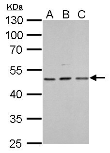
- HDAC3 Polyclonal Antibody detects HDAC3 protein by Western blot analysis. A. 30 µg A431 whole cell lysate/extract. B. 30 µg HeLa whole cell lysate/extract. C. 30 µg A375 whole cell lysate/extract.10 % SDS-PAGE. HDAC3 Polyclonal Antibody (Product # PA5-29026) dilution: 1:1,000.
- Submitted by
- Invitrogen Antibodies (provider)
- Main image

- Experimental details
- Western Blot analysis of HDAC3 was performed by separating 30 µg of various whole cell extracts by 10% SDS-PAGE. Proteins were transferred to a membrane and probed with a HDAC3 Polyclonal Antibody (Product # PA5-29026) at a dilution of 1:1000 and a HRP-conjugated anti-rabbit IgG secondary antibody.

- Submitted by
- Invitrogen Antibodies (provider)
- Main image

- Experimental details
- Western Blot using HDAC3 Polyclonal Antibody (Product # PA5-29026). Various whole cell extracts (30 µg) were separated by 10% SDS-PAGE, and the membrane was blotted with HDAC3 Polyclonal Antibody (Product # PA5-29026) diluted at 1:1,000. The HRP-conjugated anti-rabbit IgG antibody was used to detect the primary antibody.

- Submitted by
- Invitrogen Antibodies (provider)
- Main image

- Experimental details
- Western Blot analysis of HDAC3 was performed by separating 30 µg of various whole cell extracts by 10% SDS-PAGE. Proteins were transferred to a membrane and probed with a HDAC3 Polyclonal Antibody (Product # PA5-29026) at a dilution of 1:500 and a HRP-conjugated anti-rabbit IgG secondary antibody.

Supportive validation
- Submitted by
- Invitrogen Antibodies (provider)
- Main image

- Experimental details
- Immunocytochemistry-Immunofluorescence analysis of HDAC3 was performed in SK-N-SH cells fixed in 4% paraformaldehyde at RT for 15 min. Green: HDAC3 Polyclonal Antibody (Product # PA5-29026) diluted at 1:400. Red: Phalloidin, a cytoskeleton marker. Scale bar = 10 µm.

- Submitted by
- Invitrogen Antibodies (provider)
- Main image

- Experimental details
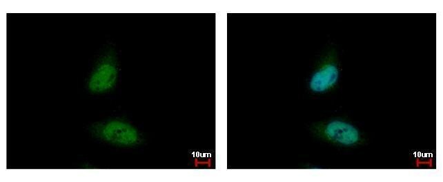
- HDAC3 Polyclonal Antibody [C3], C-term detects HDAC3 protein at nucleus by immunofluorescent analysis. Sample: HeLa cells were fixed in 4% paraformaldehyde at RT for 15 min. Green: HDAC3 protein stained by HDAC3 Polyclonal Antibody [C3], C-term (Product # PA5-29026) diluted at 1:500. Blue: Hoechst 33342 staining.
Supportive validation
- Submitted by
- Invitrogen Antibodies (provider)
- Main image

- Experimental details
- Immunohistochemical analysis of paraffin-embedded SW480 xenograft, using HDAC3 (Product # PA5-29026) antibody at 1:500 dilution. Antigen Retrieval: EDTA based buffer, pH 8.0, 15 min.

Supportive validation
- Submitted by
- Invitrogen Antibodies (provider)
- Main image

- Experimental details
- HDAC3 antibody immunoprecipitates HDAC3 protein-DNA in ChIP experiments. ChIP Sample: 293T whole cell lysate/extract. A: 5 µg preimmune rabbit IgG. B: 5 µg of HDAC3 antibody (Product # PA5-29026). The precipitated DNA was detected by PCR with primer set targeting to p21 promoter.

Supportive validation
- Submitted by
- Invitrogen Antibodies (provider)
- Main image

- Experimental details
- HDAC3 antibody immunoprecipitates HDAC3 protein in IP experiments. IP Sample: 1,000 µg 293T whole cell lysate/extract A. 50 µg 293T whole cell lysate/extract B. Control with 2 µg of preimmune rabbit IgG C. Immunoprecipitation of HDAC3 protein by 2 µg of HDAC3 antibody (Product # PA5-29026) 10% SDS-PAGE The immunoprecipitated HDAC3 protein was detected by HDAC3 antibody (Product # PA5-29026) diluted at 1:1,000.
