 Explore
 Validate
 Learn
Learn Western blot
 Immunocytochemistry
Antibody data
- Antibody Data
- Antigen structure
- References [0]
- Comments [0]
- Validations
- Western blot [1]
- Immunoprecipitation [1]
- Immunohistochemistry [3]
Submit
Validation data
Reference
Comment
Report error
- Product number
- NBP2-29906 - Provider product page

- Provider
- Novus Biologicals
- Product name
- Rabbit Polyclonal ARF1 Antibody
- Antibody type
- Polyclonal
- Description
- Immunogen affinity purified.
- Reactivity
- Human, Mouse, Canine
- Host
- Rabbit
- Isotype
- IgG
- Vial size
- 100ug
- Concentration
- 1 mg/ml
- Storage
- Store at -20C. Avoid freeze-thaw cycles.
No comments: Submit comment
Supportive validation
- Submitted by
- Novus Biologicals (provider)
- Main image

- Experimental details
- Western Blot: ARF1 Antibody [NBP2-29906] - Analysis of 25ug of various whole cell lysates per well onto a 4-20% Tris-HCl polyacrylamide gel.

Supportive validation
- Submitted by
- Novus Biologicals (provider)
- Main image

- Experimental details
- Immunoprecipitation: ARF1 Antibody [NBP2-29906] - Analysis of Arf1 was performed on HeLa cells. Antigen-antibody complexes were formed by incubating 500ug of HeLa whole cell lysate with 2ug of an Arf1 polyclonal antibody overnight on a rocking platform at 4C. The immune complexes were captured on 50ul Protein A/G Plus Agarose, washed extensively, and eluted with 5X Lane Marker Reducing Sample Buffer. Samples, including the HeLa cell lysate as a positive control (left lane), were resolved on a 4-20% Tris-HCl polyacrylamide gel, transferred to a nitrocellulose membrane, and blocked with 5% BSA/TBS-0.1%Tween for at least 1 hour. The membrane was probed with a Arf1 polyclonal antibody at a dilution of 1:1000 overnight rotating at 4C, washed in TBST, and probed with Clean-blot IP detection reagent at a dilution of 1:2500 for at least 1 hour. Chemiluminescent detection was performed using SuperSignal West Dura.

Supportive validation
- Submitted by
- Novus Biologicals (provider)
- Main image

- Experimental details
- Immunohistochemistry-Paraffin: ARF1 Antibody [NBP2-29906] - Analysis of showing staining in the cytoplasm of human colon carcinoma (right) compared to a negative control without primary antibody (left).

- Submitted by
- Novus Biologicals (provider)
- Main image
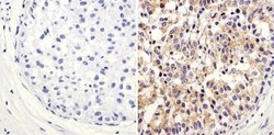
- Experimental details
- Immunohistochemistry-Paraffin: ARF1 Antibody [NBP2-29906] - Analysis showing staining in the cytoplasm of human breast carcinoma (right) compared to a negative control without primary antibody (left).

- Submitted by
- Novus Biologicals (provider)
- Main image

- Experimental details
- Immunohistochemistry-Paraffin: ARF1 Antibody [NBP2-29906] - Analysis showing staining in the cytoplasm of mouse colon tissue (right) compared to a negative control without primary antibody (left).
