Antibody data
- Antibody Data
- Antigen structure
- References [4]
- Comments [0]
- Validations
- Western blot [5]
- Immunocytochemistry [2]
- Other assay [1]
Submit
Validation data
Reference
Comment
Report error
- Product number
- PA5-29495 - Provider product page

- Provider
- Invitrogen Antibodies
- Product name
- ATP Citrate Lyase Polyclonal Antibody
- Antibody type
- Polyclonal
- Antigen
- Recombinant protein fragment
- Description
- Recommended positive controls: HeLa, Raji, mouse brain, PC-12, Rat-2. Predicted reactivity: Mouse (97%), Rat (97%), Xenopus laevis (91%), Pig (98%), Chicken (95%), Sheep (99%), Bovine (99%). Store product as a concentrated solution. Centrifuge briefly prior to opening the vial.
- Reactivity
- Human, Mouse, Rat
- Host
- Rabbit
- Isotype
- IgG
- Vial size
- 100 µL
- Concentration
- 1 mg/mL
- Storage
- Store at 4°C short term. For long term storage, store at -20°C, avoiding freeze/thaw cycles.
Submitted references BCAA Supplementation in Mice with Diet-induced Obesity Alters the Metabolome Without Impairing Glucose Homeostasis.
Dysregulated transmethylation leading to hepatocellular carcinoma compromises redox homeostasis and glucose formation.
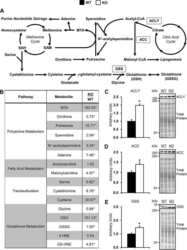
Glycine N-methyltransferase deletion in mice diverts carbon flux from gluconeogenesis to pathways that utilize excess methionine cycle intermediates.
Extended Multiplexing of Tandem Mass Tags (TMT) Labeling Reveals Age and High Fat Diet Specific Proteome Changes in Mouse Epididymal Adipose Tissue.
Lee J, Vijayakumar A, White PJ, Xu Y, Ilkayeva O, Lynch CJ, Newgard CB, Kahn BB
Endocrinology 2021 Jul 1;162(7)
Endocrinology 2021 Jul 1;162(7)
Dysregulated transmethylation leading to hepatocellular carcinoma compromises redox homeostasis and glucose formation.
Hughey CC, James FD, Wang Z, Goelzer M, Wasserman DH
Molecular metabolism 2019 May;23:1-13
Molecular metabolism 2019 May;23:1-13
Glycine N-methyltransferase deletion in mice diverts carbon flux from gluconeogenesis to pathways that utilize excess methionine cycle intermediates.
Hughey CC, Trefts E, Bracy DP, James FD, Donahue EP, Wasserman DH
The Journal of biological chemistry 2018 Jul 27;293(30):11944-11954
The Journal of biological chemistry 2018 Jul 27;293(30):11944-11954
Extended Multiplexing of Tandem Mass Tags (TMT) Labeling Reveals Age and High Fat Diet Specific Proteome Changes in Mouse Epididymal Adipose Tissue.
Plubell DL, Wilmarth PA, Zhao Y, Fenton AM, Minnier J, Reddy AP, Klimek J, Yang X, David LL, Pamir N
Molecular & cellular proteomics : MCP 2017 May;16(5):873-890
Molecular & cellular proteomics : MCP 2017 May;16(5):873-890
No comments: Submit comment
Supportive validation
- Submitted by
- Invitrogen Antibodies (provider)
- Main image

- Experimental details
- Western blot analysis was performed on whole cell extracts (30 µg lysate) of A549 (Lane 1), HeLa (Lane 2), PC-3 (Lane 3), SK-OV-3 (Lane 4), A-431 (Lane 5), Jurkat (Lane 6), Raji (Lane 7), PC-12 (Lane 8), NIH/3T3 (Lane 9) and COS-7 (Lane 10). The blot was probed with Anti-ATP Citrate Lyase Polyclonal Antibody (Product # PA5-29495, 1:1000 dilution) and detected by chemiluminescence using Goat anti-Rabbit IgG (H+L) Superclonal™ Secondary Antibody, HRP conjugate (Product # A27036, 0.25 µg/ml, 1:4000 dilution). A 120 kDa band corresponding to ATP Citrate Lyase was detected across the cell lines tested.
- Submitted by
- Invitrogen Antibodies (provider)
- Main image

- Experimental details
- Western Blot using ATP Citrate Lyase Polyclonal Antibody (Product # PA5-29495). Sample (30 µg of whole cell lysate). Lane A: HeLa. Lane B: Raji. 7.5% SDS PAGE. ATP Citrate Lyase Polyclonal Antibody (Product # PA5-29495) diluted at 1:1,000. The HRP-conjugated anti-rabbit IgG antibody was used to detect the primary antibody.
- Submitted by
- Invitrogen Antibodies (provider)
- Main image

- Experimental details
- ATP Citrate Lyase Polyclonal Antibody detects ACLY protein by western blot analysis. A. 30 µg PC-12 whole cell lysate/extract. B. 30 µg Rat2 whole cell lysate/extract.5% SDS-PAGE. ATP Citrate Lyase Polyclonal Antibody (Product # PA5-29495) dilution: 1:1,000. The HRP-conjugated anti-rabbit IgG antibody was used to detect the primary antibody.
- Submitted by
- Invitrogen Antibodies (provider)
- Main image

- Experimental details
- Western Blot using ATP Citrate Lyase Polyclonal Antibody (Product # PA5-29495). Sample (50 µg of whole cell lysate). Lane A: mouse brain. 5% SDS PAGE. ATP Citrate Lyase Polyclonal Antibody (Product # PA5-29495) diluted at 1:2,000. The HRP-conjugated anti-rabbit IgG antibody was used to detect the primary antibody.
- Submitted by
- Invitrogen Antibodies (provider)
- Main image

- Experimental details
- Knockdown of ATP Citrate Lyase was achieved by transfecting A549 cells with ATP Citrate Lyase specific siRNAs (Silencer® select Product # s916). Western blot analysis (Fig. a) was performed using whole cell extracts from the ATP Citrate Lyase knockdown cells (lane 3), non-specific scrambled siRNA transfected cells (lane 2) and untransfected cells (lane 1). The blots were probed with ATP Citrate Lyase Polyclonal Antibody (Product # PA5-29495, 1:1000 dilution) and Goat anti-Rabbit IgG (H+L) Superclonal™ Secondary Antibody, HRP conjugate (Product # A27036, 0.25 µg/ml, 1:4000 dilution). Densitometric analysis of this western blot is shown in histogram (Fig. b). Decrease in signal upon siRNA mediated knock down confirms that antibody is specific to ATP Citrate Lyase.
Supportive validation
- Submitted by
- Invitrogen Antibodies (provider)
- Main image

- Experimental details
- Immunocytochemistry-Immunofluorescence analysis of ATP Citrate Lyase was performed in HeLa cells fixed in ice cold MeOH for 5 min. Green: ATP Citrate Lyase Polyclonal Antibody (Product # PA5 29495) diluted at 1:200. Blue: Hoechst 33342 staining.
- Submitted by
- Invitrogen Antibodies (provider)
- Main image

- Experimental details
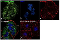
- Immunofluorescence analysis of ATP Citrate Lyase was performed using 70% confluent log phase A549 cells. The cells were fixed with 4% paraformaldehyde for 10 minutes, permeabilized with 0.1% Triton™ X-100 for 15 minutes, and blocked with 1% BSA for 1 hour at room temperature. The cells were labeled with ATP Citrate Lyase Polyclonal Antibody (Product # PA5-29495) at 5 µg/mL in 0.1% BSA, incubated at 4 degree Celsius overnight and then labeled with Goat anti-Rabbit IgG (H+L) Superclonal™ Secondary Antibody, Alexa Fluor® 488 conjugate (Product # A27034) at a dilution of 1:2000 for 45 minutes at room temperature (Panel a: green). Nuclei (Panel b: blue) were stained with ProLong™ Diamond Antifade Mountant with DAPI (Product # P36962). F-actin (Panel c: red) was stained with Rhodamine Phalloidin (Product # R415). Panel d represents the merged image showing cytoplasmic localization. Panel e represents control cells with no primary antibody to assess background. The images were captured at 60X magnification.
Supportive validation
- Submitted by
- Invitrogen Antibodies (provider)
- Main image

- Experimental details
- NULL
 Explore
 Validate
 Learn
Learn Western blot