 Explore
 Validate
 Learn
Learn10566-1-AP
antibody from Invitrogen Antibodies
Targeting: GYS1
GSY, GYS
 Western blot Immunocytochemistry
 Immunoprecipitation Immunohistochemistry Flow cytometry Other assay
Antibody data
- Antibody Data
- Antigen structure
- References [0]
- Comments [0]
- Validations
- Western blot [3]
- Immunocytochemistry [2]
- Immunohistochemistry [5]
- Flow cytometry [1]
- Other assay [1]
Submit
Validation data
Reference
Comment
Report error
- Product number
- 10566-1-AP - Provider product page

- Provider
- Invitrogen Antibodies
- Product name
- GYS1 Polyclonal Antibody
- Antibody type
- Polyclonal
- Antigen
- Other
- Description
- Immunogen sequence: RAIFATQRQ SFPPVCTHNM LDDSSDPILT TIRRIGLFNS SADRVKVIFH PEFLSSTSPL LPVDYEEFVR GCHLGVFPSY YEPWGYTPAE CTVMGIPSIS TNLSGFGCFM EEHIADPSAY GIYILDRRFR SLDDSCSQLT SFLYSFCQQS RRQRIIQRNR TERLSDLLDW KYLGRYYMSA RHMALSKAFP EHFTYEPNEA DAAQGYRYPR PASVPPSPSL SRHSSPHQSE DEEDPRNGPL EEDGERYDED EEAAKDRRNI RAPEWPRRAS CTSSTSGSKR NSVDTATSSS LSTPSEPLSP TSSLGEERN (430-737 aa encoded by BC007688)
- Reactivity
- Human, Mouse, Rat
- Host
- Rabbit
- Isotype
- IgG
- Vial size
- 150 µL
- Concentration
- 0.36 mg/mL
- Storage
- -20°C
No comments: Submit comment
Supportive validation
- Submitted by
- Invitrogen Antibodies (provider)
- Main image
- Experimental details
- K-562 cells were subjected to SDS PAGE followed by western blot with 10566-1-AP (GYS1 antibody) at dilution of 1:500 incubated at room temperature for 1.5 hours.

- Submitted by
- Invitrogen Antibodies (provider)
- Main image

- Experimental details
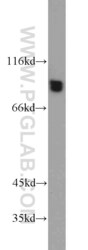
- HeLa cells were subjected to SDS PAGE followed by western blot with 10566-1-AP (GYS1 antibody) at dilution of 1:0 incubated at room temperature for 1.5 hours.
- Submitted by
- Invitrogen Antibodies (provider)
- Main image

- Experimental details
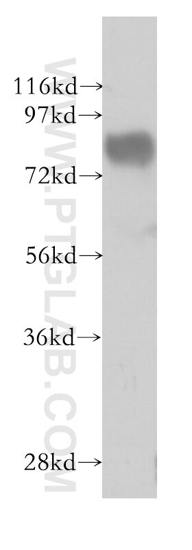
- Mouse skeletal muscle tissue were subjected to SDS PAGE followed by western blot with 10566-1-AP (GYS1 antibody) at dilution of 1:2000 incubated at room temperature for 1.5 hours.

Supportive validation
- Submitted by
- Invitrogen Antibodies (provider)
- Main image

- Experimental details
- Immunofluorescent analysis of ( -20°C Ethanol ) fixed HepG2 cells using 10566-1-AP (GYS1 antibody) at dilution of 1:50 and Alexa Fluor 488-conjugated AffiniPure Goat Anti-Rabbit IGG (H+L).

- Submitted by
- Invitrogen Antibodies (provider)
- Main image

- Experimental details
- Immunofluorescent analysis of HepG2 cells, using GYS1 antibody 10566-1-AP at 1:50 dilution and Rhodamine-labeled goat anti-rabbit IgG (red). Blue pseudocolor = DAPI (fluorescent DNA dye).

Supportive validation
- Submitted by
- Invitrogen Antibodies (provider)
- Main image

- Experimental details
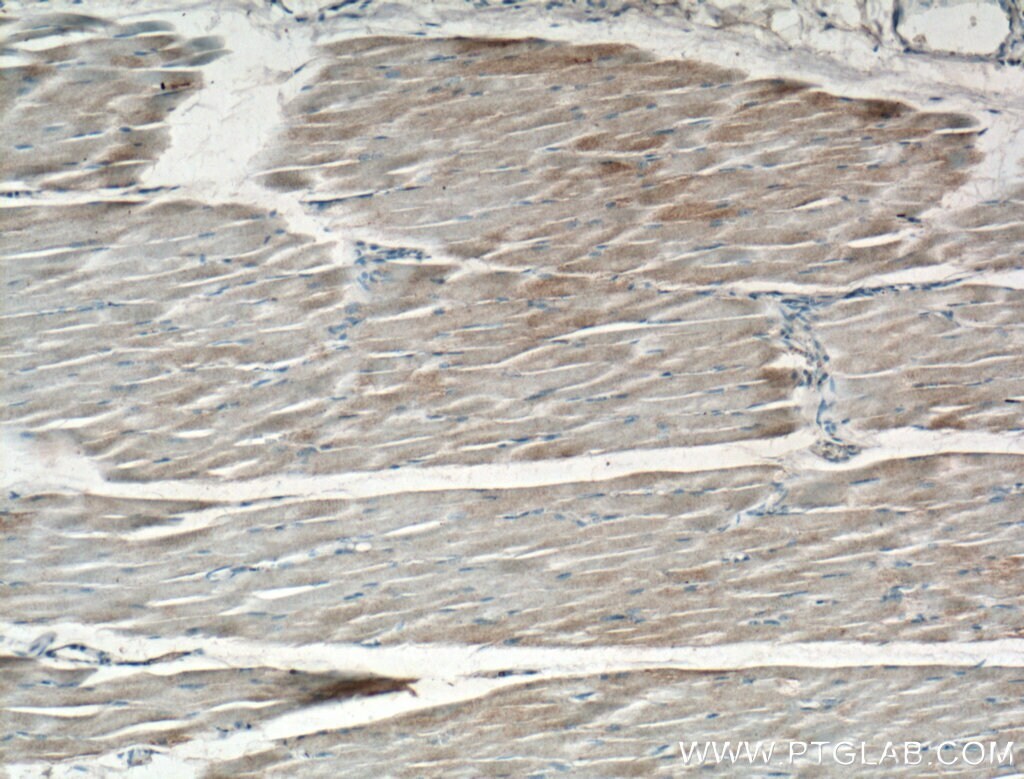
- Immunohistochemistry of paraffin-embedded human skeletal muscle tissue slide using 10566-1-AP (GYS1 antibody) at dilution of 1:200 (under 10x lens).
- Submitted by
- Invitrogen Antibodies (provider)
- Main image
- Experimental details
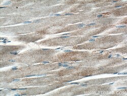
- Immunohistochemistry of paraffin-embedded human skeletal muscle tissue slide using 10566-1-AP (GYS1 antibody) at dilution of 1:200 (under 40x lens).

- Submitted by
- Invitrogen Antibodies (provider)
- Main image
- Experimental details
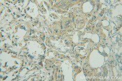
- Immunohistochemistry of paraffin-embedded human prostate cancer using 10566-1-AP (GYS1 antibody) at dilution of 1:50 (under 25x lens).

- Submitted by
- Invitrogen Antibodies (provider)
- Main image

- Experimental details
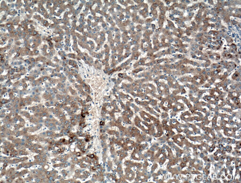
- Immunohistochemistry of paraffin-embedded human liver tissue slide using 10566-1-AP ( GYS1 antibody) at dilution of 1:200 (under 10x lens).
- Submitted by
- Invitrogen Antibodies (provider)
- Main image

- Experimental details
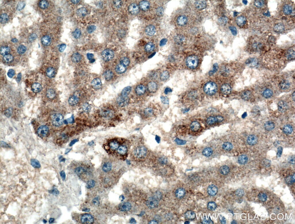
- Immunohistochemistry of paraffin-embedded human liver tissue slide using 10566-1-AP ( GYS1 antibody) at dilution of 1:200 (under 40x lens).
Supportive validation
- Submitted by
- Invitrogen Antibodies (provider)
- Main image

- Experimental details
- 1X10^6 HepG2 cells were stained with 0.2ug GYS1 antibody (10566-1-AP, red) and control antibody (blue). Fixed with 90% MeOH blocked with 3% BSA (30 min). Alexa Fluor 488-conjugated AffiniPure Goat Anti-Rabbit IGG (H+L) with dilution 1:1000.

Supportive validation
- Submitted by
- Invitrogen Antibodies (provider)
- Main image
- Experimental details
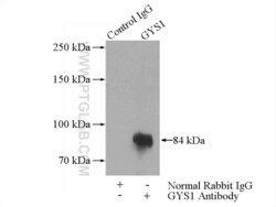
- IP result of anti-GYS1 (IP:10566-1-AP, 4ug; Detection:10566-1-AP 1:300) with mouse skeletal muscle tissue lysate 2200ug.
