Explore
 Validate
 Learn
Learn Western blot
Antibody data
- Antibody Data
- Antigen structure
- References [2]
- Comments [0]
- Validations
- Western blot [3]
- Immunocytochemistry [3]
- Immunohistochemistry [2]
- Other assay [1]
Submit
Validation data
Reference
Comment
Report error
- Product number
- MA1-22000 - Provider product page

- Provider
- Invitrogen Antibodies
- Product name
- Fibrillarin Monoclonal Antibody (38F3)
- Antibody type
- Monoclonal
- Antigen
- Other
- Description
- Store product as a concentrated solution. Centrifuge briefly prior to opening the vial.
- Reactivity
- Human, Mouse, Rat, Drosophila, Yeast
- Host
- Mouse
- Isotype
- IgG
- Antibody clone number
- 38F3
- Vial size
- 250 µL
- Concentration
- Conc. Not Determined
- Storage
- Store at 4°C short term. For long term storage, store at -20°C, avoiding freeze/thaw cycles.
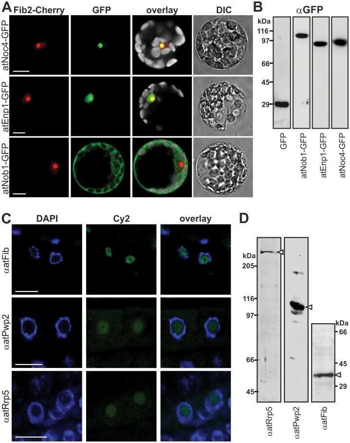
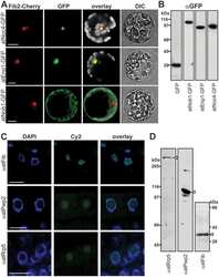
Submitted references Tau aggregates are RNA-protein assemblies that mislocalize multiple nuclear speckle components.
40S ribosome biogenesis co-factors are essential for gametophyte and embryo development.
Lester E, Ooi FK, Bakkar N, Ayers J, Woerman AL, Wheeler J, Bowser R, Carlson GA, Prusiner SB, Parker R
Neuron 2021 May 19;109(10):1675-1691.e9
Neuron 2021 May 19;109(10):1675-1691.e9
40S ribosome biogenesis co-factors are essential for gametophyte and embryo development.
Missbach S, Weis BL, Martin R, Simm S, Bohnsack MT, Schleiff E
PloS one 2013;8(1):e54084
PloS one 2013;8(1):e54084
No comments: Submit comment
Supportive validation
- Submitted by
- Invitrogen Antibodies (provider)
- Main image
- Experimental details
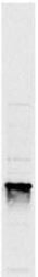
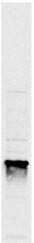
- Western blot analysis of Fibrillarin in yeast protein extract. Samples were probed with a Fibrillarin monoclonal antibody (Product # MA1-22000). The image shows a single band at ~34kDa.

- Submitted by
- Invitrogen Antibodies (provider)
- Main image

- Experimental details
- Western Blot using Fibrillarin Monoclonal Antibody (38F3) (Product # MA1-22000). Blot of yeast protein extract stained with the Fibrillarin Monoclonal Antibody (38F3) (Product # MA1-22000) anti-Gibrillarin anitbody, stained a single band at ~34 kDa.

Supportive validation
- Submitted by
- Invitrogen Antibodies (provider)
- Main image
- Experimental details
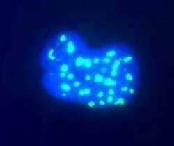
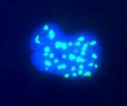
- Immunocytochemistry analysis of fibrillarin. High magnification view of human Hek293 cell nuclei stained with Fibrillarin Monoclonal Antibody (38F3) (Product # MA1-22000) (green), counterstained with a fluorescent DNA probe (blue). Nuclear DNA is revealed with Hoechst dye (blue). Cultures were processed using our standard fixat.

- Submitted by
- Invitrogen Antibodies (provider)
- Main image
- Experimental details
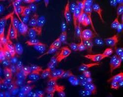
- Immunocytochemistry analysis of Human neuroblastoma line SH-SY5Y stained with Fibrillarin Monoclonal Antibody (38F3) (Product # MA1-22000) (green) and with chicken antibody to neurofilament NF-H (red) and counterstained with a fluorescent DNA probe (blue). Nuclear DNA is revealed with Hoechst dye (blue). The NF-H antibody was used at a dilution of 1:100,000 and the fibrillarin monoclonal at 1:1,000.
Supportive validation
- Submitted by
- Invitrogen Antibodies (provider)
- Main image

- Experimental details
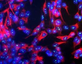
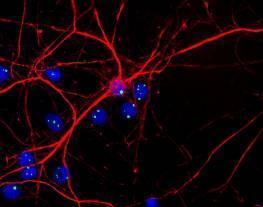
- Immunohistochemistry of rat neurons and glial stained with Fibrillarin Monoclonal Antibody (38F3) (Product # MA1-22000) (green) and with chicken antibody to neurofilament NF-H (red). Cells were counterstained with a fluorescent DNA probe (blue). Nuclear DNA is revealed with Hoechst dye (blue).

- Submitted by
- Invitrogen Antibodies (provider)
- Main image

- Experimental details
- Immunohistochemistry of rat neurons and glial stained with Fibrillarin Monoclonal Antibody (38F3) (Product # MA1-22000) (green) and with chicken antibody to neurofilament NF-H (red). Cells were counterstained with a fluorescent DNA probe (blue). Nuclear DNA is revealed with Hoechst dye (blue).
Supportive validation
- Submitted by
- Invitrogen Antibodies (provider)
- Main image
- Experimental details
- NULL