 Explore
 Validate
 Learn
Learn Western blot
Antibody data
- Antibody Data
- Antigen structure
- References [2]
- Comments [0]
- Validations
- Western blot [2]
- Immunocytochemistry [1]
Submit
Validation data
Reference
Comment
Report error
- Product number
- AF3398 - Provider product page

- Provider
- R&D Systems
- Product name
- Human/Mouse/Rat Catalase Antibody
- Antibody type
- Polyclonal
- Description
- Antigen Affinity-purified. Detects human, mouse and rat Catalase in Western blots.
- Reactivity
- Human, Mouse, Rat
- Host
- Goat
- Conjugate
- Unconjugated
- Antigen sequence
P04040- Isotype
- IgG
- Vial size
- 100 ug
- Concentration
- LYOPH
- Storage
- Use a manual defrost freezer and avoid repeated freeze-thaw cycles. 12 months from date of receipt, -20 to -70 °C as supplied. 1 month, 2 to 8 °C under sterile conditions after reconstitution. 6 months, -20 to -70 °C under sterile conditions after reconstitution.
Submitted references A critical role of autophagy in antileukemia/lymphoma effects of APO866, an inhibitor of NAD biosynthesis.
PKG inhibits TCF signaling in colon cancer cells by blocking beta-catenin expression and activating FOXO4.
Ginet V, Puyal J, Rummel C, Aubry D, Breton C, Cloux AJ, Majjigapu SR, Sordat B, Vogel P, Bruzzone S, Nencioni A, Duchosal MA, Nahimana A
Autophagy 2014 Apr;10(4):603-17
Autophagy 2014 Apr;10(4):603-17
PKG inhibits TCF signaling in colon cancer cells by blocking beta-catenin expression and activating FOXO4.
Kwon IK, Wang R, Thangaraju M, Shuang H, Liu K, Dashwood R, Dulin N, Ganapathy V, Browning DD
Oncogene 2010 Jun 10;29(23):3423-34
Oncogene 2010 Jun 10;29(23):3423-34
No comments: Submit comment
Supportive validation
- Submitted by
- R&D Systems (provider)
- Main image

- Experimental details
- Detection of Human/Mouse/Rat Catalase by Western Blot. Western blot shows lysates of Jurkat human acute T cell leukemia cell line, Raji human Burkitt's lymphoma cell line, HeLa human cervical epithelial carcinoma cell line, NIH-3T3 mouse embryonic fibroblast cell line, A20 mouse B cell lymphoma cell line, and Rat-2 rat embryonic fibroblast cell line. PVDF membrane was probed with 0.5 µg/mL of Goat Anti-Human/Mouse/Rat Catalase Antigen Affinity-purified Polyclonal Antibody (Catalog # AF3398) followed by HRP-conjugated Anti-Goat IgG Secondary Antibody (Catalog # HAF109). A specific band was detected for Catalase at approximately 64 kDa (as indicated). This experiment was conducted using Immunoblot Buffer Group 2.

- Submitted by
- R&D Systems (provider)
- Main image

- Experimental details
- Detection of Human Catalase by Simple WesternTM. Simple Western lane view shows lysates of Jurkat human acute T cell leukemia cell line and Raji human Burkitt's lymphoma cell line, loaded at 0.2 mg/mL. A specific band was detected for Catalase at approximately 62 kDa (as indicated) using 5 µg/mL of Goat Anti-Human/Mouse/Rat Catalase Antigen Affinity-purified Polyclonal Antibody (Catalog # AF3398) followed by 1:50 dilution of HRP-conjugated Anti-Goat IgG Secondary Antibody (Catalog # HAF109). This experiment was conducted under reducing conditions and using the 12-230 kDa separation system.

Supportive validation
- Submitted by
- R&D Systems (provider)
- Main image
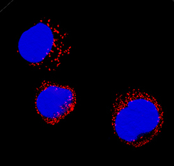
- Experimental details
- Catalase in HL-60 Human Cell Line. Catalase was detected in immersion fixed HL-60 human acute promyelocytic leukemia cell line using Goat Anti-Human/Mouse/Rat Catalase Antigen Affinity-purified Polyclonal Antibody (Catalog # AF3398) at 1.7 µg/mL for 3 hours at room temperature. Cells were stained using the NorthernLights™ 557-conjugated Anti-Goat IgG Secondary Antibody (red; Catalog # NL001) and counterstained with DAPI (blue). Specific staining was localized to peroxisomes. View our protocol for Fluorescent ICC Staining of Non-adherent Cells.
