 Explore
 Validate
 Learn
Learn Western blot
Antibody data
- Antibody Data
- Antigen structure
- References [1]
- Comments [0]
- Validations
- Western blot [1]
- Immunocytochemistry [1]
- Immunohistochemistry [1]
- Flow cytometry [1]
Submit
Validation data
Reference
Comment
Report error
- Product number
- AF7579 - Provider product page

- Provider
- R&D Systems
- Product name
- Human CD7 Antibody
- Antibody type
- Polyclonal
- Description
- Immunogen affinity purified. Detects human CD7 in direct ELISAs and Western blots. In direct ELISAs, less than 1% cross-reactivity with recombinant mouse CD7 is observed.
- Reactivity
- Human
- Host
- Sheep
- Conjugate
- Unconjugated
- Antigen sequence
P09564- Isotype
- IgG
- Vial size
- 100 ug
- Concentration
- LYOPH
- Storage
- Use a manual defrost freezer and avoid repeated freeze-thaw cycles. 12 months from date of receipt, -20 to -70 °C as supplied. 1 month, 2 to 8 °C under sterile conditions after reconstitution. 6 months, -20 to -70 °C under sterile conditions after reconstitution.
Submitted references T-cell functionality testing is highly relevant to developing novel immuno-tracers monitoring T cells in the context of immunotherapies and revealed CD7 as an attractive target.
Mayer KE, Mall S, Yusufi N, Gosmann D, Steiger K, Russelli L, Bianchi HO, Audehm S, Wagner R, Bräunlein E, Stelzl A, Bassermann F, Weichert W, Weber W, Schwaiger M, D'Alessandria C, Krackhardt AM
Theranostics 2018;8(21):6070-6087
Theranostics 2018;8(21):6070-6087
No comments: Submit comment
Supportive validation
- Submitted by
- R&D Systems (provider)
- Main image
- Experimental details
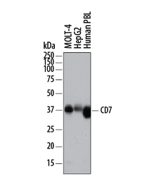
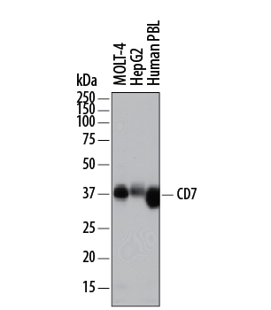
- Detection of Human CD7 by Western Blot. Western blot shows lysates of MOLT-4 human acute lymphoblastic leukemia cell line, HepG2 human hepatocellular carcinoma cell line, and human peripheral blood lymphocytes (PBL). PVDF membrane was probed with 0.5 µg/mL of Sheep Anti-Human CD7 Antigen Affinity-purified Polyclonal Antibody (Catalog # AF7579) followed by HRP-conjugated Anti-Sheep IgG Secondary Antibody (Catalog # HAF016). A specific band was detected for CD7 at approximately 35-40 kDa (as indicated). This experiment was conducted under reducing conditions and using Immunoblot Buffer Group 1.
Supportive validation
- Submitted by
- R&D Systems (provider)
- Main image
- Experimental details
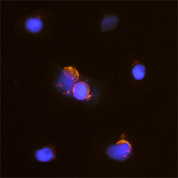
- CD7 in Human PBMCs. CD7 was detected in immersion fixed human peripheral blood mononuclear cells (PBMCs) using Sheep Anti-Human CD7 Antigen Affinity-purified Polyclonal Antibody (Catalog # AF7579) at 15 µg/mL for 3 hours at room temperature. Cells were stained using the Northern-Lights™ 557-conjugated Anti-Sheep IgG Secondary Antibody (red; Catalog # NL010) and counterstained with DAPI (blue). Specific staining was localized to cytoplasm and plasma membrane. View our protocol for Fluorescent ICC Staining of Cells on Coverslips.

Supportive validation
- Submitted by
- R&D Systems (provider)
- Main image
- Experimental details
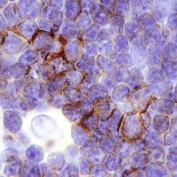
- CD7 in Human Thymus. CD7 was detected in immersion fixed paraffin-embedded sections of human thymus using Sheep Anti-Human CD7 Antigen Affinity-purified Polyclonal Antibody (Catalog # AF7579) at 3 µg/mL overnight at 4 °C. Before incubation with the primary antibody, tissue was subjected to heat-induced epitope retrieval using Antigen Retrieval Reagent-Basic (Catalog # CTS013). Tissue was stained using the Anti-Sheep HRP-DAB Cell & Tissue Staining Kit (brown; Catalog # CTS019) and counterstained with hematoxylin (blue). Specific staining was localized to the plasma membrane. View our protocol for Chromogenic IHC Staining of Paraffin-embedded Tissue Sections.

Supportive validation
- Submitted by
- R&D Systems (provider)
- Main image
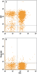
- Experimental details
- Detection of CD7 in Human PBMCs by Flow Cytometry. Human peripheral blood mononuclear cells (PBMCs) were stained with Mouse Anti-Human CD3 epsilon APC-conjugated Monoclonal Antibody (Catalog # FAB100A) and either (A) Sheep Anti-Human CD7 Antigen Affinity-purified Polyclonal Antibody (Catalog # AF7579) or (B) Sheep IgG Control (Catalog # 5-001-A) followed by Phycoerythrin-conjugated Anti-Sheep IgG Secondary Antibody (Catalog # F0126).
