 Explore
 Validate
 Learn
Learn Western blot
 ELISA
ELISAAntibody data
- Antibody Data
- Antigen structure
- References [1]
- Comments [0]
- Validations
- Western blot [1]
- Immunohistochemistry [2]
- Flow cytometry [1]
Submit
Validation data
Reference
Comment
Report error
- Product number
- AP17900PU-N - Provider product page

- Provider
- OriGene
- Product name
- Bim (BCL2L11) (Center) rabbit polyclonal antibody
- Antibody type
- Polyclonal
- Description
- Bim (BCL2L11) (Center) rabbit polyclonal antibody
- Host
- Rabbit
- Conjugate
- Unconjugated
- Epitope
- BCL2L11
- Antibody clone number
- NULL
- Vial size
- 400 µl
- Concentration
- Lot specific
Submitted references miRNA-221 and miRNA-222 induce apoptosis via the KIT/AKT signalling pathway in gastrointestinal stromal tumours.
Ihle MA, Trautmann M, Kuenstlinger H, Huss S, Heydt C, Fassunke J, Wardelmann E, Bauer S, Schildhaus HU, Buettner R, Merkelbach-Bruse S
Molecular oncology 2015 Aug;9(7):1421-33
Molecular oncology 2015 Aug;9(7):1421-33
No comments: Submit comment
Supportive validation
- Submitted by
- OriGene (provider)
- Main image

- Experimental details
- Western blot analysis of BCL2L11 Antibody (Center) in K562, HL-60 cell line lysates (35ug/lane). BCL2L11 (arrow) was detected using the purified Pab.
- Validation comment
- WB

Supportive validation
- Submitted by
- OriGene (provider)
- Main image

- Experimental details
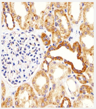
- Immunohistochemical analysis of paraffin-embedded H. kidney section using BCL2L11 Antibody (Center) (?). Antibody was diluted at 1/25 dilution. A undiluted biotinylated goat polyvalent antibody was used as the secondary, followed by DAB staining.
- Validation comment
- IHC
- Submitted by
- OriGene (provider)
- Main image
- Experimental details
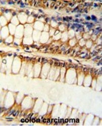
- Formalin-fixed and paraffin-embedded human colon carcinoma reacted with BCL2L11 Antibody (Center), which was peroxidase-conjugated to the secondary antibody, followed by DAB staining. This data demonstrates the use of this antibody for immunohistochemistry; clinical relevance has not been evaluated.
- Validation comment
- IHC

Supportive validation
- Submitted by
- OriGene (provider)
- Main image
- Experimental details
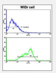
- Flow cytometric analysis of WiDr cells using BCL2L11 Antibody (Center)(bottom histogram) compared to a negative control cell (top histogram). FITC-conjugated goat-anti-rabbit secondary antibodies were used for the analysis.
- Validation comment
- FC
