Explore
 Validate
 Learn
Learn Western blot
 Immunocytochemistry
Antibody data
- Antibody Data
- Antigen structure
- References [7]
- Comments [0]
- Validations
- Western blot [1]
- Immunohistochemistry [1]
- Flow cytometry [1]
Submit
Validation data
Reference
Comment
Report error
- Product number
- V2258 - Provider product page

- Provider
- NSJ Bioreagents
- Product name
- CD43 Antibody
- Antibody type
- Monoclonal
- Antigen
- Myeloblastic KG1 cells were used as the immunogen.
- Description
- Protein G purified antibody
- Reactivity
- Human
- Host
- Mouse
- Conjugate
- Unconjugated
- Antibody clone number
- DF-T1
- Vial size
- 20 µg, 100 µg (with sodium azide), 100 µg (without sodium azide), 7 ml (prediluted for IHC use, if applicable)
- Storage
- Store the CD43 antibody at 2-8°C (with azide) or aliquot and store at -20°C or colder (without azide).
Submitted references BCL10 expression and localization in ocular adnexa MALT lymphomas: a comparative cytogenetic and immunohistochemical study.
Extranodal natural killer/T cell lymphomas with extranasal disease in non-endemic regions are disseminated or have nasal primary: a study of 84 cases from India.
Carbohydrate-mediated phagocytic recognition of early apoptotic cells undergoing transient capping of CD43 glycoprotein.
Expression of CD43 in human microglia and its downregulation in Alzheimer's disease.
Human thymic epithelial cells express an endogenous lectin, galectin-1, which binds to core 2 O-glycans on thymocytes and T lymphoblastoid cells.
T cell lines characterize events in the pathogenesis of the Wiskott-Aldrich syndrome.
Molecule detected in formalin fixed tissue by antibodies MT1, DF-T1, and L60 (Leu-22) corresponds to CD43 antigen.
Cerrone M, Collina F, De Chiara A, Corazzelli G, Curcio MP, De Renzo A, Russo F, Cantile M, Staibano S, Strianese D, Tranfa F, Botti G, De Rosa G, Franco R
Histology and histopathology 2014 Jan;29(1):77-87
Histology and histopathology 2014 Jan;29(1):77-87
Extranodal natural killer/T cell lymphomas with extranasal disease in non-endemic regions are disseminated or have nasal primary: a study of 84 cases from India.
Shet T, Suryawanshi P, Epari S, Sengar M, Rangarajan V, Menon H, Laskar S
Leukemia & lymphoma 2014 Dec;55(12):2748-53
Leukemia & lymphoma 2014 Dec;55(12):2748-53
Carbohydrate-mediated phagocytic recognition of early apoptotic cells undergoing transient capping of CD43 glycoprotein.
Eda S, Yamanaka M, Beppu M
The Journal of biological chemistry 2004 Feb 13;279(7):5967-74
The Journal of biological chemistry 2004 Feb 13;279(7):5967-74
Expression of CD43 in human microglia and its downregulation in Alzheimer's disease.
Matsuo A, Walker DG, Terai K, McGeer PL
Journal of neuroimmunology 1996 Dec;71(1-2):81-6
Journal of neuroimmunology 1996 Dec;71(1-2):81-6
Human thymic epithelial cells express an endogenous lectin, galectin-1, which binds to core 2 O-glycans on thymocytes and T lymphoblastoid cells.
Baum LG, Pang M, Perillo NL, Wu T, Delegeane A, Uittenbogaart CH, Fukuda M, Seilhamer JJ
The Journal of experimental medicine 1995 Mar 1;181(3):877-87
The Journal of experimental medicine 1995 Mar 1;181(3):877-87
T cell lines characterize events in the pathogenesis of the Wiskott-Aldrich syndrome.
Molina IJ, Kenney DM, Rosen FS, Remold-O'Donnell E
The Journal of experimental medicine 1992 Sep 1;176(3):867-74
The Journal of experimental medicine 1992 Sep 1;176(3):867-74
Molecule detected in formalin fixed tissue by antibodies MT1, DF-T1, and L60 (Leu-22) corresponds to CD43 antigen.
Stross WP, Warnke RA, Flavell DJ, Flavell SU, Simmons D, Gatter KC, Mason DY
Journal of clinical pathology 1989 Sep;42(9):953-61
Journal of clinical pathology 1989 Sep;42(9):953-61
No comments: Submit comment
Supportive validation
- Submitted by
- NSJ Bioreagents (provider)
- Main image

- Experimental details
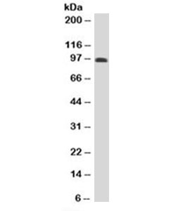
- Western blot testing of human spleen lysate with CD43 antibody (clone DF-T1). Predicted molecular weight 45-115 kDa depending on glycosylation level.
Supportive validation
- Submitted by
- NSJ Bioreagents (provider)
- Main image
- Experimental details
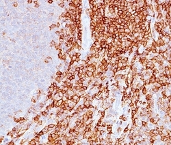
- IHC staining of spleen with CD43 antibody (DF-T1).

Supportive validation
- Submitted by
- NSJ Bioreagents (provider)
- Main image
- Experimental details
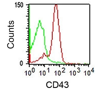
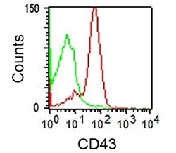
- FACS staining of human lymphocytes using DF-T1 mAb (red) and isotype control antibody.