Explore
 Validate
 Learn
Learn Immunocytochemistry
Antibody data
- Antibody Data
- Antigen structure
- References [1]
- Comments [0]
- Validations
- Immunocytochemistry [1]
- Flow cytometry [1]
Submit
Validation data
Reference
Comment
Report error
- Product number
- MAB2038 - Provider product page

- Provider
- R&D Systems
- Product name
- Human CD43 Antibody
- Antibody type
- Monoclonal
- Description
- Protein A or G purified from hybridoma culture supernatant. Detects human CD43. Stains human CD43-transfected cells but not irrelevant transfectants.
- Reactivity
- Human
- Host
- Mouse
- Conjugate
- Unconjugated
- Antigen sequence
P16150- Isotype
- IgG
- Antibody clone number
- 290111
- Vial size
- 100 ug
- Concentration
- LYOPH
- Storage
- Use a manual defrost freezer and avoid repeated freeze-thaw cycles. 12 months from date of receipt, -20 to -70 °C as supplied. 1 month, 2 to 8 °C under sterile conditions after reconstitution. 6 months, -20 to -70 °C under sterile conditions after reconstitution.
Submitted references An essential role of syntaxin 3 protein for granule exocytosis and secretion of IL-1α, IL-1β, IL-12b, and CCL4 from differentiated HL-60 cells.
Naegelen I, Plançon S, Nicot N, Kaoma T, Muller A, Vallar L, Tschirhart EJ, Bréchard S
Journal of leukocyte biology 2015 Mar;97(3):557-71
Journal of leukocyte biology 2015 Mar;97(3):557-71
No comments: Submit comment
Supportive validation
- Submitted by
- R&D Systems (provider)
- Main image

- Experimental details
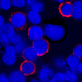
- CD43 in Human PBMCs. CD43 was detected in immersion fixed human peripheral blood mononuclear cells (PBMCs) using Mouse Anti-Human CD43 Monoclonal Antibody (Catalog # MAB2038) at 25 µg/mL for 3 hours at room temperature. Cells were stained using the NorthernLights™ 557-conjugated Anti-Mouse IgG Secondary Antibody (red; Catalog # NL007) and counterstained with DAPI (blue). Specific staining was localized to plasma membrane. View our protocol for Fluorescent ICC Staining of Cells on Coverslips.
Supportive validation
- Submitted by
- R&D Systems (provider)
- Main image
- Experimental details
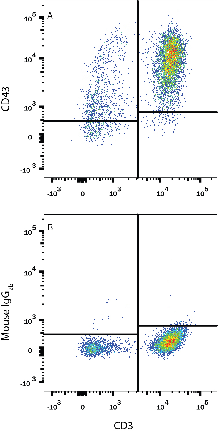
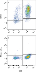
- Detection of CD43 in Human PBMCs by Flow Cytometry. Human peripheral blood mononuclear cells (PBMCs) were stained with Mouse Anti-Human CD3 epsilon APC-conjugated Monoclonal Antibody (Catalog # FAB100A) and either (A) Mouse Anti-Human CD43 Monoclonal Antibody (Catalog # MAB2038) or (B) Mouse IgG2B Flow Cytometry Isotype Control (Catalog # MAB0041) followed by Phycoerythrin-conjugated Anti-Hamster IgG Secondary Antibody. View our protocol for Staining Membrane-associated Proteins.