 Explore
 Validate
 Learn
Learn Western blot
Antibody data
- Antibody Data
- Antigen structure
- References [4]
- Comments [0]
- Validations
- Western blot [4]
- Immunocytochemistry [1]
- Immunohistochemistry [2]
- Other assay [3]
Submit
Validation data
Reference
Comment
Report error
- Product number
- PA5-21297 - Provider product page

- Provider
- Invitrogen Antibodies
- Product name
- GAD65 Polyclonal Antibody
- Antibody type
- Polyclonal
- Antigen
- Synthetic peptide
- Description
- Recommended positive controls: Mouse brain, rat brain.
- Concentration
- 1 mg/mL
Submitted references Effects of electroacupuncture on pain sensation in a rat model of hyperalgesia with nicotine dependence.
Region-Dependent Modulation of Neural Plasticity in Limbic Structures Early after Traumatic Brain Injury.
Multiple microRNAs within the 14q32 cluster target the mRNAs of major type 1 diabetes autoantigens IA-2, IA-2β, and GAD65.
Region-specific effects of repeated ketamine administration on the presynaptic GABAergic neurochemistry in rat brain.
Wang SJ, Zhang YP, Candiotti KA
Neural regeneration research 2022 Apr;17(4):905-910
Neural regeneration research 2022 Apr;17(4):905-910
Region-Dependent Modulation of Neural Plasticity in Limbic Structures Early after Traumatic Brain Injury.
Hoffman AN, Watson S, Fanselow MS, Hovda DA, Giza C
Neurotrauma reports 2021;2(1):200-213
Neurotrauma reports 2021;2(1):200-213
Multiple microRNAs within the 14q32 cluster target the mRNAs of major type 1 diabetes autoantigens IA-2, IA-2β, and GAD65.
Abuhatzira L, Xu H, Tahhan G, Boulougoura A, Schäffer AA, Notkins AL
FASEB journal : official publication of the Federation of American Societies for Experimental Biology 2015 Oct;29(10):4374-83
FASEB journal : official publication of the Federation of American Societies for Experimental Biology 2015 Oct;29(10):4374-83
Region-specific effects of repeated ketamine administration on the presynaptic GABAergic neurochemistry in rat brain.
Boczek T, Lisek M, Ferenc B, Wiktorska M, Ivchevska I, Zylinska L
Neurochemistry international 2015 Dec;91:13-25
Neurochemistry international 2015 Dec;91:13-25
No comments: Submit comment
Supportive validation
- Submitted by
- Invitrogen Antibodies (provider)
- Main image
- Experimental details
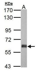
- Western blot analysis of GAD65 using 10 µg of rat brain lysate. Samples were loaded onto a 7.5% SDS-PAGE gel and probed with a GAD65 polyclonal antibody (Product # PA5-21297) at a dilution of 1:10,000.

- Submitted by
- Invitrogen Antibodies (provider)
- Main image

- Experimental details
- Western blot analysis of GAD65 using 20 µg of mouse brain lysate. Samples were loaded onto a 7.5% SDS-PAGE gel and probed with a GAD65 polyclonal antibody (Product # PA5-21297) at a dilution of 1:10,000.

- Submitted by
- Invitrogen Antibodies (provider)
- Main image
- Experimental details
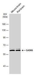
- Western Blot analysis of GAD65 was performed by separating 50 µg of Various tissue extracts by 7.5% SDS-PAGE. Proteins were transferred to a membrane and probed with a GAD65 Polyclonal Antibody (Product # PA5-21297) at a dilution of 1:5000. The HRP-conjugated anti-rabbit IgG antibody was used to detect the primary antibody.

- Submitted by
- Invitrogen Antibodies (provider)
- Main image
- Experimental details
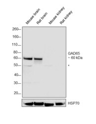
- Western blot was performed using Anti-GAD65 Polyclonal Antibody, (Product # PA5-21297) and a 60 kDa band corresponding to GAD65 was observed in the tissues tested except Mouse and Rat kidney along with an uncharacterized band (*) at ~50 kDa. Whole cell extracts (30 µg lysate) of Mouse brain (Lane 1), Rat brain (Lane 2), Mouse kidney (Lane 3), and Rat kidney (Lane 4) were electrophoresed using Novex® NuPAGE® 4-12 % Bis-Tris gel (Product # NP0321BOX). Resolved proteins were then transferred onto a nitrocellulose membrane (Product # IB23001) by iBlot® 2 Dry Blotting System (Product # IB21001). The blot was probed with the primary antibody (1:10000 dilution) and detected by chemiluminescence Goat anti-Rabbit IgG (H+L), Superclonal™ Recombinant Secondary Antibody, HRP conjugate (Product # A27036, 1:4000 dilution) using the iBright FL 1000 (Product # A32752). Chemiluminescent detection was performed using Novex® ECL Chemiluminescent Substrate Reagent Kit (Product # WP20005).

Supportive validation
- Submitted by
- Invitrogen Antibodies (provider)
- Main image

- Experimental details
- Immunocytochemistry-Immunofluorescence analysis of GAD65 was performed in Cultured Rat E18 primary cortical neuron, DIV 8. Cells fixed in 4% paraformaldehyde at RT for 15 min. Red: GAD65 Polyclonal Antibody (Product # PA5-21297) diluted at 1:250. Blue: Fluoroshield with DAPI.

Supportive validation
- Submitted by
- Invitrogen Antibodies (provider)
- Main image

- Experimental details
- Immunohistochemistry (Frozen) analysis of GAD65 was performed in frozen sectioned adult mouse retina tissue using GAD65 Polyclonal Antibody (Product # PA5-21297) at a dilution of 1:250 (Green). Red: Protein kinase C alpha staining. Blue: Fluoroshield with DAPI.

- Submitted by
- Invitrogen Antibodies (provider)
- Main image
- Experimental details
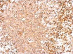
- GAD65 Polyclonal Antibody detects GAD2 protein at cytosol on RT2 xenograft by immunohistochemical analysis. Sample: Paraffin-embedded RT2 xenograft. GAD65 Polyclonal Antibody (Product # PA5-21297) dilution: 1:500. Antigen Retrieval: EDTA based buffer, pH 8.0, 15 min.

Supportive validation
- Submitted by
- Invitrogen Antibodies (provider)
- Main image

- Experimental details
- FIG. 4. Alterations in gamma-aminobutyric acid (GABA) synthesis-related proteins early after lateral fluid percussion injury (FPI). In the ipsilateral basolateral amygdala (BLA), GAD67 was increased at 24 h and decreased in the contralateral BLA at 48 h after FPI (E) . Both GAD65 and GAD67 were significantly increased in the ipsilateral dorsal hippocampus (DH) 6 h after injury (B,F) . GAD67 was also increased in the ipsilateral ventral hippocampus (VH) 6 h after FPI, where GAD65 decreased from 6 h to 24 h following FPI. (G) . In the medial prefrontal cortex (PFC), GAD67 was increased bilaterally at 24 h following injury, and significantly decreased from 48 h to 7 days in contralateral PFC. (H) . Data are represented as mean +- SEM (standard error of the mean); *** p < 0.001, ** p < 0.01, * p < 0.05, relative to sham; # p < 0.05, ## p < 0.01 across injury time-points within each blot.

- Submitted by
- Invitrogen Antibodies (provider)
- Main image
- Experimental details
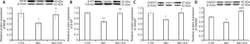
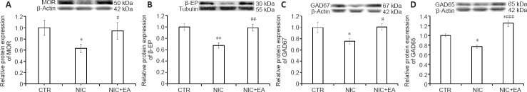
- Figure 4 Electroacupuncture effects on MOR, beta-EP, GAD67, and GAD65 protein expression in the spinal cord of nicotine-induced hyperalgesic rats . (A-D) Western blot analysis of MOR (A), beta-EP (B), GAD67 (C), and GAD65 (D) in the spinal cord of rats in the three groups. Data (mean +- SEM) are presented as relative values to control levels (CTR: n = 6, NIC: n = 6, NIC + EA: n = 6). * P < 0.05, ** P < 0.01, vs . CTR group; # P < 0.05, ## P < 0.01, ### P < 0.001, vs . NIC group (one-way analysis of variance followed by Newman-Keuls multiple comparison test). CTR: Age-matched control group; NIC: nicotine-exposed group; NIC + EA: nicotine-exposed/electroacupuncture-treated group. GAD65: glutamic acid decarboxylase 65; GAD67: glutamic acid decarboxylase 67; MOR: mu-opioid receptor; beta-EP: beta-endorphin.
- Submitted by
- Invitrogen Antibodies (provider)
- Main image

- Experimental details
- Figure 5 Electroacupuncture effects on MOR, beta-EP, GAD67, and GAD65 protein expression in the PAG of nicotine-induced hyperalgesic rats . (A-D) Western blot analysis of MOR (A), beta-EP (B), GAD67 (C), and GAD65 (D) in the PAG of rats in the three groups. Data (mean +- SEM) are presented as relative values to control levels (CTR: n = 6, NIC: n = 6, NIC + EA: n = 6). * P < 0.05, ** P < 0.01, vs . CTR group; # P < 0.05, ## P < 0.01, vs . NIC group (one-way analysis of variance followed by Newman-Keuls multiple comparison test). CTR: Age-matched control group; NIC: nicotine-exposed group; NIC + EA: nicotine-exposed/electroacupuncture-treated group. GAD65: glutamic acid decarboxylase 65; GAD67: glutamic acid decarboxylase 67; MOR: mu-opioid receptor; PAG: periaqueductal gray; beta-EP: beta-endorphin.
