Antibody data
- Antibody Data
- Antigen structure
- References [6]
- Comments [0]
- Validations
- Western blot [2]
- Immunohistochemistry [4]
- Other assay [4]
Submit
Validation data
Reference
Comment
Report error
- Product number
- PA5-16867 - Provider product page

- Provider
- Invitrogen Antibodies
- Product name
- Claudin 3 Polyclonal Antibody
- Antibody type
- Polyclonal
- Antigen
- Synthetic peptide
- Description
- PA5-16867 targets Claudin 3 in IHC (P) and WB applications and shows reactivity with Human, mouse, and Rat samples.
Submitted references Inhibition of interferon-signalling halts cancer-associated fibroblast-dependent protection of breast cancer cells from chemotherapy.
Claudin expression profile in flat wart and cutaneous squamous cell carcinoma in epidermodysplasia verruciformis.
MyD88 regulates a prolonged adaptation response to environmental dust exposure-induced lung disease.
TLR3 deficiency exacerbates the loss of epithelial barrier function during genital tract Chlamydia muridarum infection.
Proximal tubular cells contain a phenotypically distinct, scattered cell population involved in tubular regeneration.
Integrin Beta 1 suppresses multilayering of a simple epithelium.
Broad RV, Jones SJ, Teske MC, Wastall LM, Hanby AM, Thorne JL, Hughes TA
British journal of cancer 2021 Mar;124(6):1110-1120
British journal of cancer 2021 Mar;124(6):1110-1120
Claudin expression profile in flat wart and cutaneous squamous cell carcinoma in epidermodysplasia verruciformis.
da Cruz Silva LL, de Oliveira WRP, Pereira NV, Halpern I, Tanabe CKD, Mattos MSG, Sotto MN
Scientific reports 2020 Jun 9;10(1):9268
Scientific reports 2020 Jun 9;10(1):9268
MyD88 regulates a prolonged adaptation response to environmental dust exposure-induced lung disease.
Johnson AN, Harkema JR, Nelson AJ, Dickinson JD, Kalil J, Duryee MJ, Thiele GM, Kumar B, Singh AB, Gaurav R, Glover SC, Tang Y, Romberger DJ, Kielian T, Poole JA
Respiratory research 2020 Apr 22;21(1):97
Respiratory research 2020 Apr 22;21(1):97
TLR3 deficiency exacerbates the loss of epithelial barrier function during genital tract Chlamydia muridarum infection.
Kumar R, Gong H, Liu L, Ramos-Solis N, Seye CI, Derbigny WA
PloS one 2019;14(1):e0207422
PloS one 2019;14(1):e0207422
Proximal tubular cells contain a phenotypically distinct, scattered cell population involved in tubular regeneration.
Smeets B, Boor P, Dijkman H, Sharma SV, Jirak P, Mooren F, Berger K, Bornemann J, Gelman IH, Floege J, van der Vlag J, Wetzels JF, Moeller MJ
The Journal of pathology 2013 Apr;229(5):645-59
The Journal of pathology 2013 Apr;229(5):645-59
Integrin Beta 1 suppresses multilayering of a simple epithelium.
Chen J, Krasnow MA
PloS one 2012;7(12):e52886
PloS one 2012;7(12):e52886
No comments: Submit comment
Supportive validation
- Submitted by
- Invitrogen Antibodies (provider)
- Main image

- Experimental details
- Western blot analysis was performed on whole cell extracts (30 µg lysate) of MCF-7 (Lane 1), PC-3 (Lane 2), LNCaP (Lane 3), and Caco-2 (Lane 4). The blots were probed with Anti-Claudin-3 Rabbit Polyclonal Antibody (Product # PA5-16867, 1-2 µg/mL) and detected by chemiluminescence using Goat anti-Rabbit IgG (H+L) Secondary Antibody, HRP conjugate (Product # G-21234, 1:5000 dilution). A 18 kDa band corresponding to Claudin-3 was observed across cell lines tested. Known quantity of protein samples were electrophoresed using Novex® NuPAGE® 12 % Bis-Tris gel (Product # NP0342BOX), XCell SureLock™ Electrophoresis System (Product # EI0002) and Novex® Sharp Pre-Stained Protein Standard (Product # LC5800). Resolved proteins were then transferred onto a nitrocellulose membrane with iBlot® 2 Dry Blotting System (Product # IB21001). The membrane was probed with the relevant primary and secondary Antibody following blocking with 5 % skimmed milk. Chemiluminescent detection was performed using Pierce™ ECL Western Blotting Substrate (Product # 32106).
- Submitted by
- Invitrogen Antibodies (provider)
- Main image

- Experimental details
- Knockdown of Claudin 3 was achieved by transfecting PC-3 with Claudin 3 specific siRNAs (Silencer® select Product # s3445, s3444). Western blot analysis (Fig. a) was performed using membrane extracts from the Claudin 3 knockdown cells (lane 3), non-specific scrambled siRNA transfected cells (lane 2) and untransfected cells (lane 1). The blots were probed with Claudin 3 Polyclonal Antibody (Product # PA5-16867, 1 µg/mL) and Goat anti-Rabbit IgG (H+L) Superclonal™ Secondary Antibody, HRP conjugate (Product # A27036, 0.25 µg/mL, 1:4000 dilution). Densitometric analysis of this western blot is shown in histogram (Fig. b). Decrease in signal upon siRNA mediated knock down confirms that antibody is specific to Claudin 3.
Supportive validation
- Submitted by
- Invitrogen Antibodies (provider)
- Main image

- Experimental details
- Formalin-fixed, paraffin-embedded human small intestine stained with Claudin3 using peroxidase-conjugate and AEC. Note membrane staining of epithelial cells
- Submitted by
- Invitrogen Antibodies (provider)
- Main image

- Experimental details
- Immunohistochemistry analysis of Claudin 3 showing staining in the membrane of paraffin-embedded human small intestine tissue (right) compared to a negative control without primary antibody (left). To expose target proteins, antigen retrieval was performed using 10mM sodium citrate (pH 6.0), microwaved for 8-15 min. Following antigen retrieval, tissues were blocked in 3% H2O2-methanol for 15 min at room temperature, washed with ddH2O and PBS, and then probed with a Claudin 3 Rabbit Polyclonal Antibody (Product # PA5-16867) diluted in 3% BSA-PBS at a dilution of 1:20 for 1 hour at 37°C in a humidified chamber. Tissues were washed extensively in PBST and detection was performed using an HRP-conjugated secondary antibody followed by colorimetric detection using a DAB kit. Tissues were counterstained with hematoxylin and dehydrated with ethanol and xylene to prep for mounting.
- Submitted by
- Invitrogen Antibodies (provider)
- Main image

- Experimental details
- Immunohistochemistry analysis of Claudin 3 showing staining in the membrane of paraffin-embedded mouse colon tissue (right) compared to a negative control without primary antibody (left). To expose target proteins, antigen retrieval was performed using 10mM sodium citrate (pH 6.0), microwaved for 8-15 min. Following antigen retrieval, tissues were blocked in 3% H2O2-methanol for 15 min at room temperature, washed with ddH2O and PBS, and then probed with a Claudin 3 Rabbit Polyclonal Antibody (Product # PA5-16867) diluted in 3% BSA-PBS at a dilution of 1:20 for 1 hour at 37°C in a humidified chamber. Tissues were washed extensively in PBST and detection was performed using an HRP-conjugated secondary antibody followed by colorimetric detection using a DAB kit. Tissues were counterstained with hematoxylin and dehydrated with ethanol and xylene to prep for mounting.
- Submitted by
- Invitrogen Antibodies (provider)
- Main image

- Experimental details
- Immunohistochemistry analysis of Claudin 3 showing staining in the membrane of paraffin-embedded mouse liver tissue (right) compared to a negative control without primary antibody (left). To expose target proteins, antigen retrieval was performed using 10mM sodium citrate (pH 6.0), microwaved for 8-15 min. Following antigen retrieval, tissues were blocked in 3% H2O2-methanol for 15 min at room temperature, washed with ddH2O and PBS, and then probed with a Claudin 3 Rabbit Polyclonal Antibody (Product # PA5-16867) diluted in 3% BSA-PBS at a dilution of 1:20 for 1 hour at 37°C in a humidified chamber. Tissues were washed extensively in PBST and detection was performed using an HRP-conjugated secondary antibody followed by colorimetric detection using a DAB kit. Tissues were counterstained with hematoxylin and dehydrated with ethanol and xylene to prep for mounting.
Supportive validation
- Submitted by
- Invitrogen Antibodies (provider)
- Main image

- Experimental details
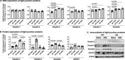
- Fig. 6 Repetitive ODE exposure increases expression of several tight junction proteins known to be upregulated in inflamed/injured lung in WT mice but not in MyD88 KO mice. WT and MyD88 KO mice were treated i.n. daily for 3 weeks with saline or ODE. Panel a, Expression of tight junction mRNA was measured by real-time quantitative PCR and are reported as fold-changes normalized to control. Panel b , Quantification of tight junction protein expression in ODE-treated mice compared to control mice as determined by the immunoblot presented in Panel c . Scatter plots demonstrate mean with standard error bars of N = 3 animals per group with 2 replicates per sample
- Submitted by
- Invitrogen Antibodies (provider)
- Main image

- Experimental details
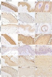
- Figure 1 Epidermodysplasia verruciformis group: claudins expression in normal skin, flat wart and cutaneous squamous cell carcinoma (cSCC). ( a-c ) Claudin-1 membranous bound staining, with diffuse distribution in all histological types. ( d-f ) Claudin-2 diffuse expression, with membranous and cytoplasmatic immunostaining. ( g-i ) Claudin-3 diffuse expression in normal skin (upper layers) and flat wart, however with focal expression in cSCC. ( j-l ) Claudin-4 diffuse distribution, with cytoplasmatic immunostaining. ( m-o ) Claudin-5 focal expression in normal skin (upper layers), but with diffuse pattern in flat wart and cSCC. Diffuse expression of claudin-7 ( p-r ) and claudin-11 ( s-u ) in all histological types, with cytoplasmatic immunostaining. Immunohistochemistry images photographed by the author in 2019 .
- Submitted by
- Invitrogen Antibodies (provider)
- Main image

- Experimental details
- Figure 2 Not epidermodysplasia verruciformis group: claudins expression in normal skin, flat wart and cutaneous squamous cell carcinoma (cSCC). ( a-c ) Claudin-1 membranous bound staining, with diffuse distribution in all histological types. ( d-f ) Claudin-2 diffuse expression, with membranous and cytoplasmatic immunostaining. ( g-i ) Claudin-3 diffuse expression in normal skin (upper layers) and flat wart, however with focal expression in cSCC, with positivity at the center of the tumor islands. (j-l) Claudin-4 diffuse expression, with membranous and cytoplasmatic immunostaining. ( m-o ) Claudin-5 focal expression in normal skin (upper layers) and flat wart, but with diffuse distribution in cSCC. Diffuse expression of claudin-7 ( p-r ) and claudin-11 ( s-u ) in all histological types, with cytoplasmatic immunostaining. Immunohistochemistry images photographed by the author in 2019 .
- Submitted by
- Invitrogen Antibodies (provider)
- Main image

- Experimental details
- Fig. 5 In primary cancers, IFNbeta1 in CAFs and MX1 in cancer cells correlate with each other and with poor survival. TMAs of tissue from 109 TNBC resections were assembled and expression of IFNbeta1 in fibroblasts, and MX1 and claudin-3 in tumour cells was determined using immunohistochemistry. a Representative images of immunohistochemistry, showing tissue scored '3' for IFNbeta in fibroblasts (left), '3' for MX1, and 'positive' for claudin-3. b The cohort was split into groups with high or low expression of IFNbeta1 in fibroblasts (left) or MX1 in tumour cells (right) using ROC analyses. Cumulative disease-free survival in the groups was compared using Kaplan-Meier analyses and log rank tests. c The cohort was split into claudin-low or claudin-high groups, based on expression levels of claudin-3 (positive or negative). The claudin-low group ( n = 49) were analysed as in b .
 Explore
 Validate
 Learn
Learn Western blot