 Explore
 Validate
 Learn
Learn Immunocytochemistry
Antibody data
- Antibody Data
- Antigen structure
- References [1]
- Comments [0]
- Validations
- Immunocytochemistry [1]
- Immunohistochemistry [1]
- Flow cytometry [1]
Submit
Validation data
Reference
Comment
Report error
- Product number
- MAB4620 - Provider product page

- Provider
- R&D Systems
- Product name
- Human Claudin-3 Antibody
- Antibody type
- Monoclonal
- Description
- Protein A or G purified from hybridoma culture supernatant. Stains human Claudin-3 transfectants but not irrelevant transfectants.
- Reactivity
- Human
- Host
- Mouse
- Conjugate
- Unconjugated
- Antigen sequence
O15551- Isotype
- IgG
- Antibody clone number
- 385021
- Vial size
- 100 ug
- Concentration
- LYOPH
- Storage
- Use a manual defrost freezer and avoid repeated freeze-thaw cycles. 12 months from date of receipt, -20 to -70 °C as supplied. 1 month, 2 to 8 °C under sterile conditions after reconstitution. 6 months, -20 to -70 °C under sterile conditions after reconstitution.
Submitted references Label-free detection and molecular profiling of exosomes with a nano-plasmonic sensor.
Im H, Shao H, Park YI, Peterson VM, Castro CM, Weissleder R, Lee H
Nature biotechnology 2014 May;32(5):490-5
Nature biotechnology 2014 May;32(5):490-5
No comments: Submit comment
Supportive validation
- Submitted by
- R&D Systems (provider)
- Main image

- Experimental details
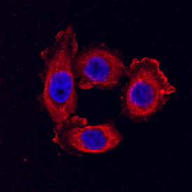
- Claudin-3 in PC-3 Human Cell Line. Claudin-3 was detected in immersion fixed PC-3 human prostate cancer cell line using Mouse Anti-Human Claudin-3 Monoclonal Antibody (Catalog # MAB4620) at 10 µg/mL for 3 hours at room temperature. Cells were stained using the NorthernLights™ 557-conjugated Anti-Mouse IgG Secondary Antibody (red; Catalog # NL007) and counterstained with DAPI (blue). Specific staining was localized to cytoplasm. View our protocol for Fluorescent ICC Staining of Cells on Coverslips.
Supportive validation
- Submitted by
- R&D Systems (provider)
- Main image

- Experimental details
- Claudin-3 in Human Colon. Claudin-3 was detected in immersion fixed paraffin-embedded sections of human colon array using Mouse Anti-Human Claudin-3 Monoclonal Antibody (Catalog # MAB4620) at 15 µg/mL overnight at 4 °C. Tissue was stained using the Anti-Mouse HRP-DAB Cell & Tissue Staining Kit (brown; Catalog # CTS002) and counterstained with hematoxylin (blue). Lower panel shows a lack of labeling if primary antibodies are omitted and tissue is stained only with secondary antibody followed by incubation with detection reagents. View our protocol for Chromogenic IHC Staining of Paraffin-embedded Tissue Sections.

Supportive validation
- Submitted by
- R&D Systems (provider)
- Main image

- Experimental details
- Detection of Claudin-3 in PC-3 Human Cell Line by Flow Cytometry. PC-3 human prostate cancer cell line was stained with Mouse Anti-Human Claudin-3 Monoclonal Antibody (Catalog # MAB4620, filled histogram) or isotype control antibody (Catalog # MAB003, open histogram), followed by Phycoerythrin-conjugated Anti-Mouse IgG F(ab')2 Secondary Antibody (Catalog # F0102B).
