Explore
 Validate
 Learn
Learn Western blot
 Immunoprecipitation
Antibody data
- Antibody Data
- Antigen structure
- References [0]
- Comments [0]
- Validations
- Western blot [6]
- Immunocytochemistry [1]
- Immunohistochemistry [4]
Submit
Validation data
Reference
Comment
Report error
- Product number
- PA5-30044 - Provider product page

- Provider
- Invitrogen Antibodies
- Product name
- GNAO1 Polyclonal Antibody
- Antibody type
- Polyclonal
- Antigen
- Recombinant protein fragment
- Description
- Recommended positive controls: U87-MG, SK-N-SH, IMR32, SK-N-AS, mouse brain, mouse lung, rat brain, rat lung. Predicted reactivity: Mouse (97%), Rat (98%), Xenopus laevis (98%), Chicken (98%), Rhesus Monkey (100%), Bovine (98%). Store product as a concentrated solution. Centrifuge briefly prior to opening the vial.
- Reactivity
- Human, Mouse, Rat
- Host
- Rabbit
- Isotype
- IgG
- Vial size
- 100 µL
- Concentration
- 1 mg/mL
- Storage
- Store at 4°C short term. For long term storage, store at -20°C, avoiding freeze/thaw cycles.
No comments: Submit comment
Supportive validation
- Submitted by
- Invitrogen Antibodies (provider)
- Main image
- Experimental details
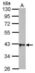
- Western blot analysis of GNAO1 using 5 µg of mouse brain lysate. Samples were loaded onto a 10% SDS-PAGE gel and probed with a GNAO1 polyclonal antibody (Product # PA5-30044) at a dilution of 1:10,000.

- Submitted by
- Invitrogen Antibodies (provider)
- Main image
- Experimental details
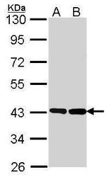
- Western blot analysis of GNAO1 using 30 µg of A) HeLa and B) NT2D1 lysate. Samples were loaded onto a 10% SDS-PAGE gel and probed with a GNAO1 polyclonal antibody (Product # PA5-30044) at a dilution of 1:1000.
- Submitted by
- Invitrogen Antibodies (provider)
- Main image

- Experimental details
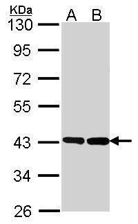
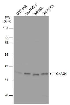
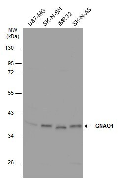
- Western Blot analysis of GNAO1 was performed by separating 30 µg of various whole cell extracts by 10% SDS-PAGE. Proteins were transferred to a membrane and probed with a GNAO1 Polyclonal Antibody (Product # PA5-30044) at a dilution of 1:1000 and a HRP-conjugated anti-rabbit IgG secondary antibody.
- Submitted by
- Invitrogen Antibodies (provider)
- Main image
- Experimental details
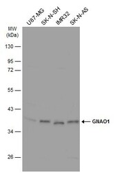
- Western Blot analysis of GNAO1 was performed by separating 30 µg of various whole cell extracts by 10% SDS-PAGE. Proteins were transferred to a membrane and probed with a GNAO1 Polyclonal Antibody (Product # PA5-30044) at a dilution of 1:1000 and a HRP-conjugated anti-rabbit IgG secondary antibody.
- Submitted by
- Invitrogen Antibodies (provider)
- Main image

- Experimental details
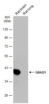
- Western blot analysis of GNAO1 was performed by separating 50 µg of various tissue extracts by 10% SDS-PAGE. Proteins were transferred to a membrane and probed with a GNAO1 Polyclonal Antibody (Product # PA5-30044) at a dilution of 1:10000. The HRP-conjugated anti-rabbit IgG antibody was used to detect the primary antibody.
- Submitted by
- Invitrogen Antibodies (provider)
- Main image
- Experimental details
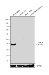
- Western blot was performed using Anti-GNAO1 Polyclonal Antibody (Product # PA5-30044) and a 40kDa band corresponding to GNAO1 was observed in Mouse Brain and not in Mouse Lung, Mouse Kidney and Mouse Skeletal Muscle which are reported negative for GNAO1 expression. Tissue extracts (30 µg lysate) of Mouse Brain (Lane 1), Mouse Lung (Lane 2), Mouse Kidney (Lane 3) and Mouse Skeletal Muscle (Lane 4) were electrophoresed using Novex® NuPAGE® 4-12 % Bis-Tris gel (Product # NP0322BOX). Resolved proteins were then transferred onto a nitrocellulose membrane (Product # IB23001) by iBlot® 2 Dry Blotting System (Product # IB21001). The blot was probed with the primary antibody (1:1000 dilution) and detected by chemiluminescence Goat Anti-Rabbit IgG Secondary Antibody, HRP conjugate (Product # A27036, 1:4000 dilution) using the iBright FL 1000 (Product # A32752). Chemiluminescent detection was performed using Novex® ECL Chemiluminescent Substrate Reagent Kit (Product # WP20005).

Supportive validation
- Submitted by
- Invitrogen Antibodies (provider)
- Main image

- Experimental details
- Immunocytochemistry-Immunofluorescence analysis of GNAO1 was performed in DIV9 rat E18 primary hippocampal neuron cells fixed in 4% paraformaldehyde at RT for 15 min. Green: GNAO1 Polyclonal Antibody (Product # PA5-30044) diluted at 1:500. Red: beta Tubulin 3/ Tuj1, stained by beta Tubulin 3/ Tuj1 antibody. Blue: Fluoroshield with DAPI.

Supportive validation
- Submitted by
- Invitrogen Antibodies (provider)
- Main image
- Experimental details
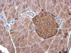
- Immunohistochemistry (Paraffin) analysis of GNAO1 was performed in paraffin-embedded mouse pancreas tissue using GNAO1 Polyclonal Antibody (Product # PA5-30044) at a dilution of 1:500.

- Submitted by
- Invitrogen Antibodies (provider)
- Main image

- Experimental details
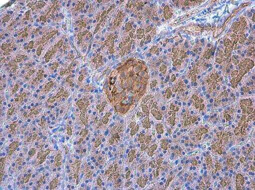
- Immunohistochemistry (Paraffin) analysis of GNAO1 was performed in paraffin-embedded rat pancreas tissue using GNAO1 Polyclonal Antibody (Product # PA5-30044) at a dilution of 1:500.
- Submitted by
- Invitrogen Antibodies (provider)
- Main image
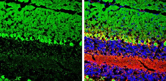
- Experimental details
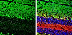
- Immunohistochemistry (Frozen) analysis of GNAO1 was performed in frozen-sectioned mouse cerebellum tissue using GNAO1 Polyclonal Antibody (Product # PA5-30044) at a dilution of 1:250 (Green). Red: NF-H, stained by NF-H antibody diluted at 1:500. Blue: Fluoroshield with DAPI.
- Submitted by
- Invitrogen Antibodies (provider)
- Main image
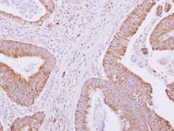
- Experimental details
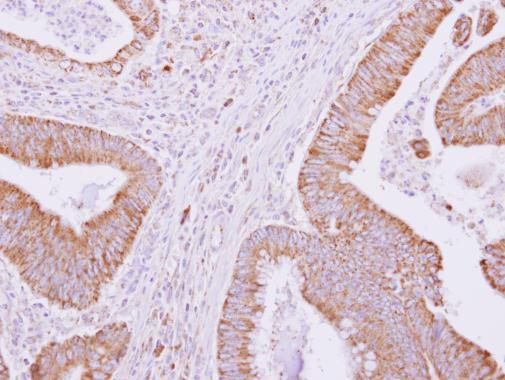
- GNAO1 Polyclonal Antibody detects GNAO1 protein at cytoplasm on human colon carcinoma by immunohistochemical analysis. Sample: Paraffin-embedded colon carcinoma. GNAO1 Polyclonal Antibody (Product # PA5-30044) dilution: 1:500. Antigen Retrieval: EDTA based buffer, pH 8.0, 15 min.