Explore
 Validate
 Learn
Learn Western blot
 Immunocytochemistry
 Immunohistochemistry
Antibody data
- Antibody Data
- Antigen structure
- References [3]
- Comments [0]
- Validations
- Immunocytochemistry [1]
- Immunohistochemistry [4]
Submit
Validation data
Reference
Comment
Report error
- Product number
- HPA000913 - Provider product page

- Provider
- Atlas Antibodies
- Proper citation
- Atlas Antibodies Cat#HPA000913, RRID:AB_1858795
- Product name
- Anti-WDR13
- Antibody type
- Polyclonal
- Reactivity
- Human
- Host
- Rabbit
- Conjugate
- Unconjugated
- Antigen sequence
GHRRSVSRGSYQLQAQMNRAVYEDRPPGSVVPTSA
AEASRAMAGDTSLSENYAFAGMYHVFDQHVDEAVP
RVRFANDDRHRLACCSLDGSISLCQLVPAPPTVLR
VLR- Isotype
- IgG
- Vial size
- 100 µl
- Storage
- Store at +4°C for short term storage. Long time storage is recommended at -20°C.
Submitted references Role of mouse Wdr13 in placental growth; a genetic evidence for lifetime body weight determination by placenta during development.
Immunofluorescence and fluorescent-protein tagging show high correlation for protein localization in mammalian cells
Lack of Wdr13 gene in mice leads to enhanced pancreatic beta cell proliferation, hyperinsulinemia and mild obesity.
Singh VP, Alex JL, Lakshmi BJ, Sailasree SP, Raj TA, Kumar S
Scientific reports 2015 Aug 26;5:13371
Scientific reports 2015 Aug 26;5:13371
Immunofluorescence and fluorescent-protein tagging show high correlation for protein localization in mammalian cells
Stadler C, Rexhepaj E, Singan V, Murphy R, Pepperkok R, Uhlén M, Simpson J, Lundberg E
Nature Methods 2013 February;10(4):315-323
Nature Methods 2013 February;10(4):315-323
Lack of Wdr13 gene in mice leads to enhanced pancreatic beta cell proliferation, hyperinsulinemia and mild obesity.
Singh VP, Lakshmi BJ, Singh S, Shah V, Goel S, Sarathi DP, Kumar S
PloS one 2012;7(6):e38685
PloS one 2012;7(6):e38685
No comments: Submit comment
Supportive validation
- Submitted by
- Atlas Antibodies (provider)
- Main image

- Experimental details
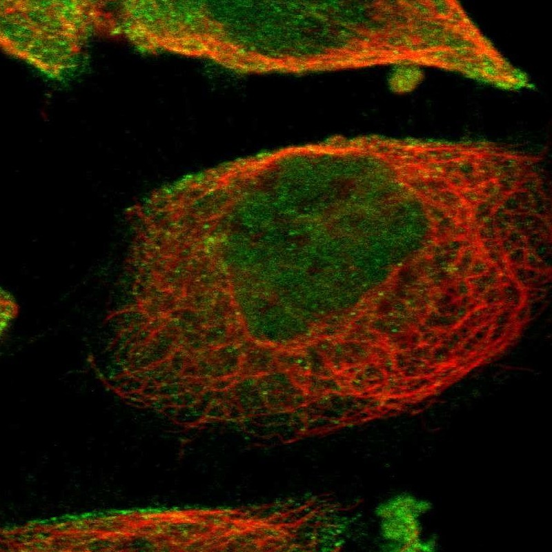
- Immunofluorescent staining of human cell line A-431 shows localization to nucleoplasm & plasma membrane.
- Sample type
- HUMAN
Enhanced validation
Supportive validation
- Submitted by
- Atlas Antibodies (provider)
- Enhanced method
- Orthogonal validation
- Main image
- Experimental details
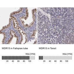
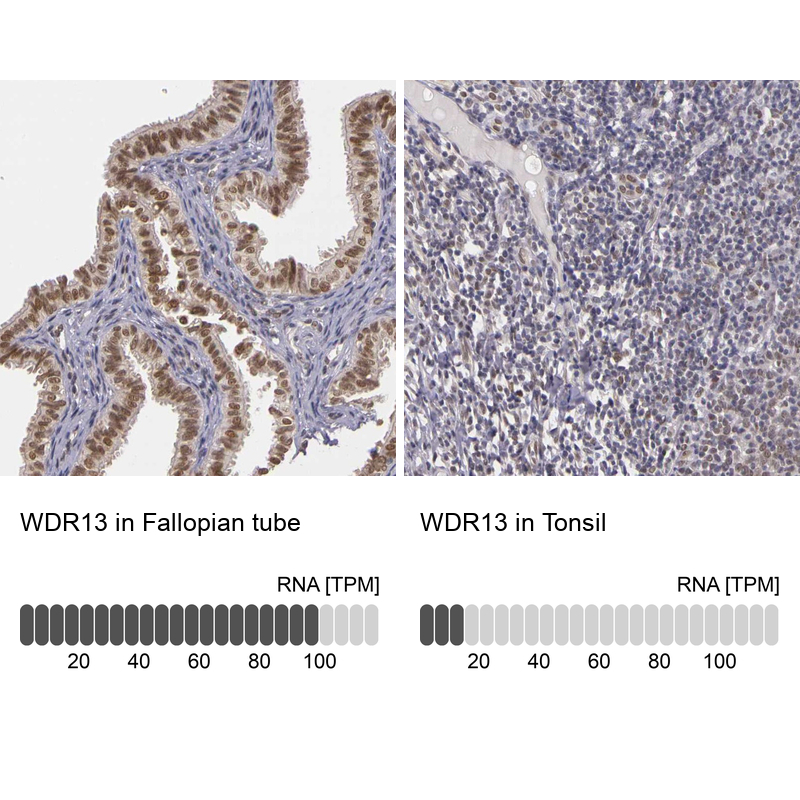
- Immunohistochemistry analysis in human fallopian tube and tonsil tissues using Anti-WDR13 antibody. Corresponding WDR13 RNA-seq data are presented for the same tissues.
- Sample type
- HUMAN
Supportive validation
- Submitted by
- Atlas Antibodies (provider)
- Main image
- Experimental details
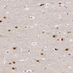
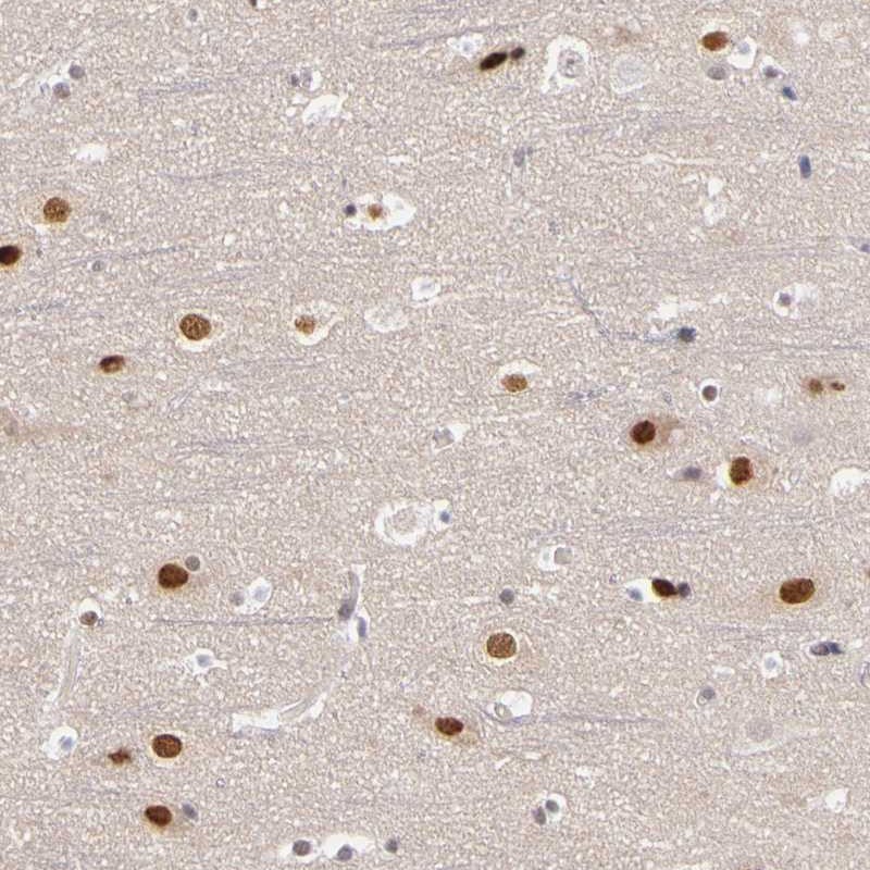
- Immunohistochemical staining of human cerebral cortex shows moderate nuclear positivity in neuronal cells.
- Submitted by
- Atlas Antibodies (provider)
- Main image
- Experimental details
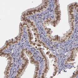
- Immunohistochemical staining of human fallopian tube shows high expression.
- Sample type
- HUMAN

- Submitted by
- Atlas Antibodies (provider)
- Main image

- Experimental details
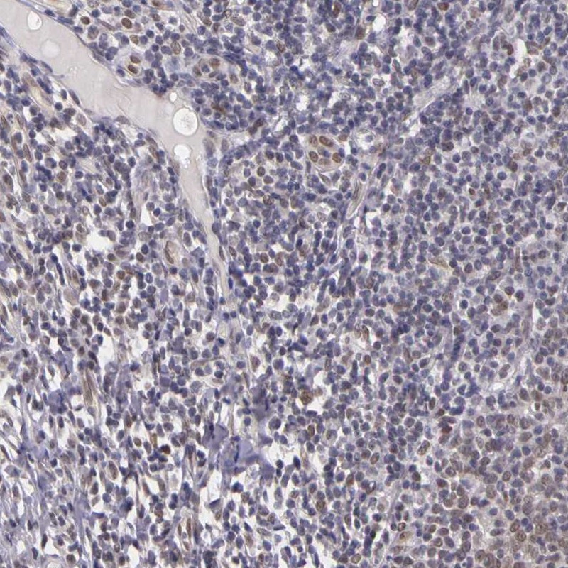
- Immunohistochemical staining of human tonsil shows low expression as expected.
- Sample type
- HUMAN