Antibody data
- Antibody Data
- Antigen structure
- References [20]
- Comments [0]
- Validations
- Western blot [2]
- Immunocytochemistry [1]
- Immunohistochemistry [1]
- Other assay [5]
Submit
Validation data
Reference
Comment
Report error
- Product number
- MA1-113 - Provider product page

- Provider
- Invitrogen Antibodies
- Product name
- MDM2 Monoclonal Antibody (IF2)
- Antibody type
- Monoclonal
- Antigen
- Other
- Description
- This antibody recognizes the ~90 kDa (apparent MW) MDM2 protein. Also recognizes isoforms at ~57 and ~74/76 kDa. This antibody was originally validated as part of a Thermo Scientific Cellomics High Content Screening Kit. The antibody sold separately may have slightly different performance and may need to be further optimized for the best results.
- Reactivity
- Human
- Host
- Mouse
- Isotype
- IgG
- Antibody clone number
- IF2
- Vial size
- 100 µg
- Concentration
- 1.0 mg/mL
- Storage
- -20° C, Avoid Freeze/Thaw Cycles
Submitted references Expression levels of FBXW7 and MDM2 E3 ubiquitin ligases and their c-Myc and p53 substrates in patients with dysplastic nevi or melanoma.
Cancer Cells Employ Nuclear Caspase-8 to Overcome the p53-Dependent G2/M Checkpoint through Cleavage of USP28.
PIDDosome-induced p53-dependent ploidy restriction facilitates hepatocarcinogenesis.
MDM2 Derived from Dedifferentiated Liposarcoma Extracellular Vesicles Induces MMP2 Production from Preadipocytes.
Dual Targeting of Aurora Kinases with AMG 900 Exhibits Potent Preclinical Activity Against Acute Myeloid Leukemia with Distinct Post-Mitotic Outcomes.
Prognostic value of HMGA2, CDK4, and JUN amplification in well-differentiated and dedifferentiated liposarcomas.
Myxoid liposarcoma with heterologous components: dedifferentiation or metaplasia? A FISH-documented and CGH-documented case report.
MDM2 and CDK4 immunohistochemical coexpression in high-grade osteosarcoma: correlation with a dedifferentiated subtype.
MDM2 and CDK4 immunohistochemistry is a valuable tool in the differential diagnosis of low-grade osteosarcomas and other primary fibro-osseous lesions of the bone.
Well-differentiated liposarcoma with low-grade osteosarcomatous component: an underrecognized variant.
Immunohistochemical analysis of MDM2 and CDK4 distinguishes low-grade osteosarcoma from benign mimics.
Similarity in genetic alterations between paired well-differentiated and dedifferentiated components of dedifferentiated liposarcoma.
Markers aiding the diagnosis of chondroid tumors: an immunohistochemical study including osteonectin, bcl-2, cox-2, actin, calponin, D2-40 (podoplanin), mdm-2, CD117 (c-kit), and YKL-40.
Lipoleiomyosarcoma of the rectosigmoid colon: a unique site for a rare variant of liposarcoma.
Immunostaining for peroxisome proliferator gamma distinguishes dedifferentiated liposarcoma from other retroperitoneal sarcomas.
Sporadic desmoid tumor. An exceptional cause of cystic pancreatic lesion.
alpha-fetoprotein expression in a dedifferentiated liposarcoma.
Palmar atypical lipomatous tumour with spindle cell features (well-differentiated spindle cell liposarcoma): a rare neoplasm arising in an unusual anatomical location.
Inflammatory malignant fibrous histiocytomas and dedifferentiated liposarcomas: histological review, genomic profile, and MDM2 and CDK4 status favour a single entity.
Most malignant fibrous histiocytomas developed in the retroperitoneum are dedifferentiated liposarcomas: a review of 25 cases initially diagnosed as malignant fibrous histiocytoma.
Mozuraitiene J, Gudleviciene Z, Vincerzevskiene I, Laurinaviciene A, Pamedys J
Oncology letters 2021 Jan;21(1):37
Oncology letters 2021 Jan;21(1):37
Cancer Cells Employ Nuclear Caspase-8 to Overcome the p53-Dependent G2/M Checkpoint through Cleavage of USP28.
Müller I, Strozyk E, Schindler S, Beissert S, Oo HZ, Sauter T, Lucarelli P, Raeth S, Hausser A, Al Nakouzi N, Fazli L, Gleave ME, Liu H, Simon HU, Walczak H, Green DR, Bartek J, Daugaard M, Kulms D
Molecular cell 2020 Mar 5;77(5):970-984.e7
Molecular cell 2020 Mar 5;77(5):970-984.e7
PIDDosome-induced p53-dependent ploidy restriction facilitates hepatocarcinogenesis.
Sladky VC, Knapp K, Szabo TG, Braun VZ, Bongiovanni L, van den Bos H, Spierings DC, Westendorp B, Curinha A, Stojakovic T, Scharnagl H, Timelthaler G, Tsuchia K, Pinter M, Semmler G, Foijer F, de Bruin A, Reiberger T, Rohr-Udilova N, Villunger A
EMBO reports 2020 Dec 3;21(12):e50893
EMBO reports 2020 Dec 3;21(12):e50893
MDM2 Derived from Dedifferentiated Liposarcoma Extracellular Vesicles Induces MMP2 Production from Preadipocytes.
Casadei L, Calore F, Braggio DA, Zewdu A, Deshmukh AA, Fadda P, Lopez G, Wabitsch M, Song C, Leight JL, Grignol VP, Lev D, Croce CM, Pollock RE
Cancer research 2019 Oct 1;79(19):4911-4922
Cancer research 2019 Oct 1;79(19):4911-4922
Dual Targeting of Aurora Kinases with AMG 900 Exhibits Potent Preclinical Activity Against Acute Myeloid Leukemia with Distinct Post-Mitotic Outcomes.
Payton M, Cheung HK, Ninniri MSS, Marinaccio C, Wayne WC, Hanestad K, Crispino JD, Juan G, Coxon A
Molecular cancer therapeutics 2018 Dec;17(12):2575-2585
Molecular cancer therapeutics 2018 Dec;17(12):2575-2585
Prognostic value of HMGA2, CDK4, and JUN amplification in well-differentiated and dedifferentiated liposarcomas.
Saâda-Bouzid E, Burel-Vandenbos F, Ranchère-Vince D, Birtwisle-Peyrottes I, Chetaille B, Bouvier C, Château MC, Peoc'h M, Battistella M, Bazin A, Gal J, Michiels JF, Coindre JM, Pedeutour F, Bianchini L
Modern pathology : an official journal of the United States and Canadian Academy of Pathology, Inc 2015 Nov;28(11):1404-14
Modern pathology : an official journal of the United States and Canadian Academy of Pathology, Inc 2015 Nov;28(11):1404-14
Myxoid liposarcoma with heterologous components: dedifferentiation or metaplasia? A FISH-documented and CGH-documented case report.
Weingertner N, Neuville A, Chibon F, Ray-Coquard I, Marcellin L, Ghnassia JP
Applied immunohistochemistry & molecular morphology : AIMM 2015 Mar;23(3):230-5
Applied immunohistochemistry & molecular morphology : AIMM 2015 Mar;23(3):230-5
MDM2 and CDK4 immunohistochemical coexpression in high-grade osteosarcoma: correlation with a dedifferentiated subtype.
Yoshida A, Ushiku T, Motoi T, Beppu Y, Fukayama M, Tsuda H, Shibata T
The American journal of surgical pathology 2012 Mar;36(3):423-31
The American journal of surgical pathology 2012 Mar;36(3):423-31
MDM2 and CDK4 immunohistochemistry is a valuable tool in the differential diagnosis of low-grade osteosarcomas and other primary fibro-osseous lesions of the bone.
Dujardin F, Binh MB, Bouvier C, Gomez-Brouchet A, Larousserie F, Muret Ad, Louis-Brennetot C, Aurias A, Coindre JM, Guillou L, Pedeutour F, Duval H, Collin C, de Pinieux G
Modern pathology : an official journal of the United States and Canadian Academy of Pathology, Inc 2011 May;24(5):624-37
Modern pathology : an official journal of the United States and Canadian Academy of Pathology, Inc 2011 May;24(5):624-37
Well-differentiated liposarcoma with low-grade osteosarcomatous component: an underrecognized variant.
Yoshida A, Ushiku T, Motoi T, Shibata T, Fukayama M, Tsuda H
The American journal of surgical pathology 2010 Sep;34(9):1361-6
The American journal of surgical pathology 2010 Sep;34(9):1361-6
Immunohistochemical analysis of MDM2 and CDK4 distinguishes low-grade osteosarcoma from benign mimics.
Yoshida A, Ushiku T, Motoi T, Shibata T, Beppu Y, Fukayama M, Tsuda H
Modern pathology : an official journal of the United States and Canadian Academy of Pathology, Inc 2010 Sep;23(9):1279-88
Modern pathology : an official journal of the United States and Canadian Academy of Pathology, Inc 2010 Sep;23(9):1279-88
Similarity in genetic alterations between paired well-differentiated and dedifferentiated components of dedifferentiated liposarcoma.
Horvai AE, DeVries S, Roy R, O'Donnell RJ, Waldman F
Modern pathology : an official journal of the United States and Canadian Academy of Pathology, Inc 2009 Nov;22(11):1477-88
Modern pathology : an official journal of the United States and Canadian Academy of Pathology, Inc 2009 Nov;22(11):1477-88
Markers aiding the diagnosis of chondroid tumors: an immunohistochemical study including osteonectin, bcl-2, cox-2, actin, calponin, D2-40 (podoplanin), mdm-2, CD117 (c-kit), and YKL-40.
Daugaard S, Christensen LH, Høgdall E
APMIS : acta pathologica, microbiologica, et immunologica Scandinavica 2009 Jul;117(7):518-25
APMIS : acta pathologica, microbiologica, et immunologica Scandinavica 2009 Jul;117(7):518-25
Lipoleiomyosarcoma of the rectosigmoid colon: a unique site for a rare variant of liposarcoma.
Nahal A, Meterissian S
American journal of clinical oncology 2009 Aug;32(4):353-5
American journal of clinical oncology 2009 Aug;32(4):353-5
Immunostaining for peroxisome proliferator gamma distinguishes dedifferentiated liposarcoma from other retroperitoneal sarcomas.
Horvai AE, Schaefer JT, Nakakura EK, O'Donnell RJ
Modern pathology : an official journal of the United States and Canadian Academy of Pathology, Inc 2008 May;21(5):517-24
Modern pathology : an official journal of the United States and Canadian Academy of Pathology, Inc 2008 May;21(5):517-24
Sporadic desmoid tumor. An exceptional cause of cystic pancreatic lesion.
Amiot A, Dokmak S, Sauvanet A, Vilgrain V, Bringuier PP, Scoazec JY, Sastre X, Ruszniewski P, Bedossa P, Couvelard A
JOP : Journal of the pancreas 2008 May 8;9(3):339-45
JOP : Journal of the pancreas 2008 May 8;9(3):339-45
alpha-fetoprotein expression in a dedifferentiated liposarcoma.
Bosco M, Allia E, Coindre JM, Odasso C, Pagani A, Pacchioni D
Virchows Archiv : an international journal of pathology 2006 Apr;448(4):517-20
Virchows Archiv : an international journal of pathology 2006 Apr;448(4):517-20
Palmar atypical lipomatous tumour with spindle cell features (well-differentiated spindle cell liposarcoma): a rare neoplasm arising in an unusual anatomical location.
Mentzel T, Toennissen J, Rütten A, Schaller J
Virchows Archiv : an international journal of pathology 2005 Mar;446(3):300-4
Virchows Archiv : an international journal of pathology 2005 Mar;446(3):300-4
Inflammatory malignant fibrous histiocytomas and dedifferentiated liposarcomas: histological review, genomic profile, and MDM2 and CDK4 status favour a single entity.
Coindre JM, Hostein I, Maire G, Derré J, Guillou L, Leroux A, Ghnassia JP, Collin F, Pedeutour F, Aurias A
The Journal of pathology 2004 Jul;203(3):822-30
The Journal of pathology 2004 Jul;203(3):822-30
Most malignant fibrous histiocytomas developed in the retroperitoneum are dedifferentiated liposarcomas: a review of 25 cases initially diagnosed as malignant fibrous histiocytoma.
Coindre JM, Mariani O, Chibon F, Mairal A, De Saint Aubain Somerhausen N, Favre-Guillevin E, Bui NB, Stoeckle E, Hostein I, Aurias A
Modern pathology : an official journal of the United States and Canadian Academy of Pathology, Inc 2003 Mar;16(3):256-62
Modern pathology : an official journal of the United States and Canadian Academy of Pathology, Inc 2003 Mar;16(3):256-62
No comments: Submit comment
Supportive validation
- Submitted by
- Invitrogen Antibodies (provider)
- Main image

- Experimental details
- Western blot analysis of MDM2 was performed by loading 20 µg of the indicated whole cell lysates and 5 µL of PageRuler Plus Prestained Protein Ladder (Product # 26619) per well onto a Novex 4-20% Tris-Glycine polyacrylamide gel (Product # WT4202BOX ). Proteins were transferred to a nitrocellulose membrane using the G2 Blotter (Product # 62288), and blocked with 5% Milk in TBST for 1 hour at room temperature. MDM2 was detected at ~57, ~75, and ~90 kDa using a MDM2 monoclonal antibody (Product # MA1-113) at a dilution of 1 µg/mL in 5% Milk in TBST overnight at 4C on a rocking platform, followed by a Goat anti-Mouse IgG (H+L) Superclonal Secondary Antibody, HRP conjugate (Product # A28177) at a dilution of 1:1000 for at least 30 minutes at room temperature. Chemiluminescent detection was performed using SuperSignal Pico substrate (Product # 34078) and the myECL Imager (Product # 62236).
- Submitted by
- Invitrogen Antibodies (provider)
- Main image
- Experimental details
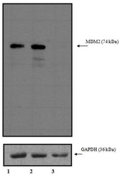
- Western blot analysis of MDM2 was performed with 10 µg of A549 cells transfected with Transfection Reagent alone (Lane 1), 100nM Non-Targeting control siRNA (Lane 2), or 100nM siRNA against MDM2 (Lane 3). Proteins were resolved using a NuPAGE® Novex 4-12% Bis-Tris Gel (Product # NP0322BOX), XCell SureLock™ Electrophoresis System (Product # EI0002), and a protein size ladder. Proteins were wet transferred to a Pierce Nitrocellulose Membrane (Product # 88025) OR Pierce PVDF Membrane (Product # 88518) and blocked with Pierce Starting Block T20 (PBS) Blocking Buffer (Product # 37539) for 1 hour at room temperature. MDM2 was detected at ~ 74 kDa using MDM2 Mouse monoclonal antibody (Product # MA1-113) diluted in Pierce Starting Block T20 (PBS) Blocking Buffer 4°C overnight on a rocking platform. Pierce Goat Anti-Mouse (Product # 31437) HRP-Conjugated Antibodies at a 1:2500 dilution were used and chemiluminescent detection was performed using Pierce Supersignal West Dura Maximum Sensitivity Substrate (Product # 37071). Relative density of the bands normalized to GAPDH (36 kDa). MDM2 Antibody (Product # MA1-113) confirms silencing of MDM2 expression.
Supportive validation
- Submitted by
- Invitrogen Antibodies (provider)
- Main image
- Experimental details
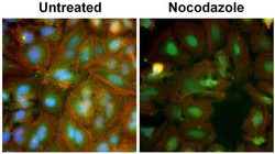
- Immunofluorescent analysis of MDM2 (green) in HeLa cells either left untreated (left panel) or treated with 50nM Nocodazole (right panel) for 16 hours. Formalin fixed cells were permeabilized with 0.1% Triton X-100 in PBS for 15 minutes at room temperature and blocked with 3% Blocker BSA (Product # 37525) for 15 minutes at room temperature. Cells were probed with a MDM2 monoclonal antibody (Product # MA1-113) at a concentration of 1 µg/mL for at least 1 hour at room temperature, washed with PBS, and incubated with a Goat anti-Mouse IgG (H+L) Superclonal Secondary Antibody, Alexa Fluor 488 conjugate (Product # A28175) at a dilution of 1:400 for 30 minutes at room temperature. Actin (red) was stained with DyLight 554 Phalloidin (Product # 21834) and nuclei (blue) were stained with Hoechst 33342 dye (Product # 62249). Images were taken on a Thermo Scientific ArrayScan or ToxInsight Instrument at 20X magnification.
Supportive validation
- Submitted by
- Invitrogen Antibodies (provider)
- Main image

- Experimental details
- Immunohistochemistry analysis of MDM2 showing staining in the cytoplasm and nucleus of paraffin-embedded human breast carcinoma (right) compared to a negative control without primary antibody (left). To expose target proteins, antigen retrieval was performed using 10mM sodium citrate (pH 6.0) and heated in a 95C water bath for 20 minutes. Following antigen retrieval, tissues were blocked in 10% goat serum in PBS for 30 minutes at room temperature and quenched with Peroxide Suppressor (Product # 35000) for 30 minutes. Tissues were then probed with a MDM2 monoclonal antibody (Product # MA1-113) at a dilution of 40 µg/mL in blocking buffer for 1 hour at room temperature. Tissues were washed extensively in PBST and detection was performed using the SuperPicture HRP Polymer Detection Kit (Product # 87-8963) and DAB substrate (Product # 34002). Tissues were counterstained with hematoxylin (Product # TA-125-MH) and dehydrated with ethanol and xylene to prep for mounting.
Supportive validation
- Submitted by
- Invitrogen Antibodies (provider)
- Main image
- Experimental details
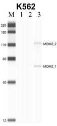
- Immunoprecipitation of MDM2 was performed in K562 cells. Antigen-antibody complexes were formed by incubating approximately 500 µg whole cell lysate with 5 to 10 µL of monoclonal MDM2 antibody (Product # MA1-113) rotating 60 min at RT. The immune complexes were captured on 625 µg of anti-mouse coated Dynabeads (Product # 11202D) and washed extensively. They were then eluted and analyzed using the Simple Western system using the same antibody as used in immunoprecipitation at a dilution of 1:25, followed by a 1:100 dilution of secondary antibody. Lane 1 is the input, lane 2 no antibody IP and lane 3 is the target specific IP. Data courtesy of the Yeo lab as part of the ENCODE project.
- Submitted by
- Invitrogen Antibodies (provider)
- Main image

- Experimental details
- NULL
- Submitted by
- Invitrogen Antibodies (provider)
- Main image
- Experimental details
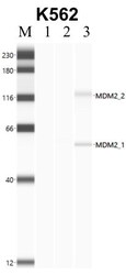
- Immunoprecipitation of MDM2 was performed in K562 cells. Antigen-antibody complexes were formed by incubating approximately 500 µg whole cell lysate with 5 to 10 µL of monoclonal MDM2 antibody (Product # MA1-113) rotating 60 min at RT. The immune complexes were captured on 625 µg of anti-mouse coated Dynabeads (Product # 11202D) and washed extensively. They were then eluted and analyzed using the Simple Western system using the same antibody as used in immunoprecipitation at a dilution of 1:25, followed by a 1:100 dilution of secondary antibody. Lane 1 is the input, lane 2 no antibody IP and lane 3 is the target specific IP. Data courtesy of the Yeo lab as part of the ENCODE project.
- Submitted by
- Invitrogen Antibodies (provider)
- Main image

- Experimental details
- Figure 6. MDM2 protein expression in cutaneous melanoma. Representative images of (A) weak MDM2 immunostaining in pT1 melanoma and (B) in pT2 melanoma, (C) moderate MDM2 immunostaining in pT3 melanoma and (D) weak MDM2immunostaining in pT4 melanoma. Marked areas indicate MDM2 staining in malignant melanocytes. Magnification, x100. pT, pathological tumor stage.
- Submitted by
- Invitrogen Antibodies (provider)
- Main image
- Experimental details
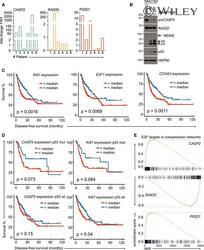
- EV4 Figure Evaluation of PIDDosome expression levels and its prognostic value in human HCC Densitometric quantification of the immunoblot in Fig 5A is shown as the fold change in tumor (T) tissue over non-tumorous (NT) tissue of the individual patients. The values for proCASP2, PIDD-C, and RAIDD were normalized to the corresponding PonceauS staining. Immunoblot of the HCC cell line SNU182 treated with DMSO or increasing concentrations of the Aurora B inhibitor ZM447439. The membrane was probed for CASP2, RAIDD, MDM2, p53, and HSP90 as loading control. Recurrence-free survival of HCC patients in the TGCA Provisional data set (LIHC) expressing high or low levels of the proliferation-associated genes Ki67 , E2F1, and CCNA2 . Disease-free survival of the TGCA HCC patients shown in Fig 5C was analyzed with respect to the p53 (mutated or wt) status. Data are divided at the median, and statistical significance was tested using a log-rank test. Butterfly plots showing the enrichment of E2F target genes in the co-expression networks of CASP2 , RAIDD, and PIDD1 in the same TCGA data set. Source data are available online for this figure.
 Explore
 Validate
 Learn
Learn Western blot
 Immunoprecipitation