Explore
 Validate
 Learn
Learn Western blot
 Immunocytochemistry
Antibody data
- Antibody Data
- Antigen structure
- References [4]
- Comments [0]
- Validations
- Western blot [7]
- Immunoprecipitation [1]
- Immunohistochemistry [2]
Submit
Validation data
Reference
Comment
Report error
- Product number
- NBP1-31330 - Provider product page

- Provider
- Novus Biologicals
- Proper citation
- Novus Cat#NBP1-31330, RRID:AB_10003781
- Product name
- Rabbit Polyclonal Cyclin A2 Antibody
- Antibody type
- Polyclonal
- Description
- Immunogen affinity purified.
- Reactivity
- Human, Mouse, Rat
- Host
- Rabbit
- Isotype
- IgG
- Vial size
- 0.1 ml
- Storage
- Aliquot and store at -20C or -80C. Avoid freeze-thaw cycles.
Submitted references Botulinum toxin type A affects the transcriptome of cell cultures derived from muscle biopsies of controls and spastic patients.
Hepatic inflammation caused by dysregulated bile acid synthesis is reversible by butyrate supplementation.
Similarities in Gene Expression Profiles during In Vitro Aging of Primary Human Embryonic Lung and Foreskin Fibroblasts.
Suppression of cell-cycle progression by Jun dimerization protein-2 (JDP2) involves downregulation of cyclin-A2.
Zanotti S, Kapetis D, Gibertini S, Salerno F, Ciusani E, Colombo C, Gronchi A, Morandi L, Mantegazza R, Molteni F, Mora M
Toxicology in vitro : an international journal published in association with BIBRA 2018 Aug;50:124-136
Toxicology in vitro : an international journal published in association with BIBRA 2018 Aug;50:124-136
Hepatic inflammation caused by dysregulated bile acid synthesis is reversible by butyrate supplementation.
Sheng L, Jena PK, Hu Y, Liu HX, Nagar N, Kalanetra KM, French SW, French SW, Mills DA, Wan YY
The Journal of pathology 2017 Dec;243(4):431-441
The Journal of pathology 2017 Dec;243(4):431-441
Similarities in Gene Expression Profiles during In Vitro Aging of Primary Human Embryonic Lung and Foreskin Fibroblasts.
Marthandan S, Priebe S, Baumgart M, Groth M, Cellerino A, Guthke R, Hemmerich P, Diekmann S
BioMed research international 2015;2015:731938
BioMed research international 2015;2015:731938
Suppression of cell-cycle progression by Jun dimerization protein-2 (JDP2) involves downregulation of cyclin-A2.
Pan J, Nakade K, Huang YC, Zhu ZW, Masuzaki S, Hasegawa H, Murata T, Yoshiki A, Yamaguchi N, Lee CH, Yang WC, Tsai EM, Obata Y, Yokoyama KK
Oncogene 2010 Nov 25;29(47):6245-56
Oncogene 2010 Nov 25;29(47):6245-56
No comments: Submit comment
Supportive validation
- Submitted by
- Novus Biologicals (provider)
- Main image
- Experimental details
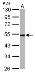
- Western Blot: Cyclin A2 Antibody [NBP1-31330] - 30 ug PC-12 whole cell lysate/extract 10% SDS-PAGE gel, antibody dilution 1:1000.

- Submitted by
- Novus Biologicals (provider)
- Main image

- Experimental details
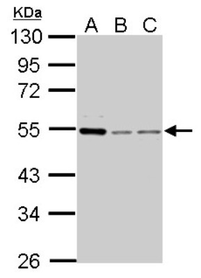
- Western Blot: Cyclin A2 Antibody [NBP1-31330] - A. 30 ug 293T whole cell lysate/extract, B. 30 ug HeLa whole cell lysate/extract, C. 30 ug A37C whole cell lysate/extract 10% SDS-PAGE gel, antibody dilution 1:1000.
- Submitted by
- Novus Biologicals (provider)
- Main image
- Experimental details
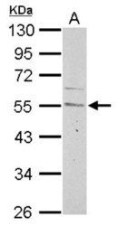
- Western Blot: Cyclin A2 Antibody [NBP1-31330] - 30 ug Neuro2A whole cell lysate/extract 10% SDS-PAGE gel, antibody dilution 1:1000.

- Submitted by
- Novus Biologicals (provider)
- Main image
- Experimental details
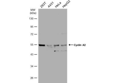
- Western Blot: Cyclin A2 Antibody [NBP1-31330] - Various whole cell extracts (30 ug) were separated by 10% SDS-PAGE, and the membrane was blotted with Cyclin A2 antibody diluted at a dilution of 1:1000.
- Submitted by
- Novus Biologicals (provider)
- Main image
- Experimental details
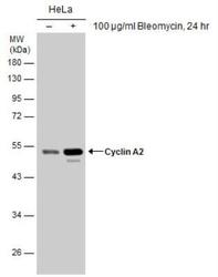
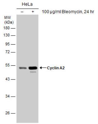
- Western Blot: Cyclin A2 Antibody [NBP1-31330] - Untreated (-) and treated (+) HeLa whole cell extracts (30 ug) were separated by 10% SDS-PAGE, and the membrane was blotted with Cyclin A2 antibody. HRP-conjugated anti-rabbit IgG antibody was used to detect the primary antibody.
- Submitted by
- Novus Biologicals (provider)
- Main image

- Experimental details
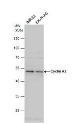
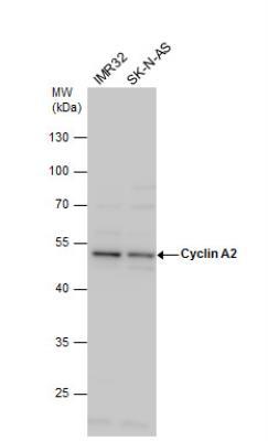
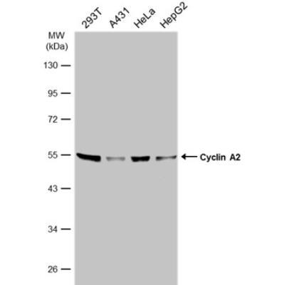
- Western Blot: Cyclin A2 Antibody [NBP1-31330] - Various whole cell extracts (30 ug) were separated by 10% SDS-PAGE, and the membrane was blotted with Cyclin A2 antibody diluted at 1:1000. The HRP-conjugated anti-rabbit IgG antibody (NBP2-19301) was used to detect the primary antibody.
- Submitted by
- Novus Biologicals (provider)
- Main image

- Experimental details
- Western Blot: Cyclin A2 Antibody [NBP1-31330] - Various whole cell extracts (30 ug) were separated by 10% SDS-PAGE, and the membrane was blotted with Cyclin A2 antibody diluted at 1:1000. The HRP-conjugated anti-rabbit IgG antibody (NBP2-19301) was used to detect the primary antibody.
Supportive validation
- Submitted by
- Novus Biologicals (provider)
- Main image

- Experimental details
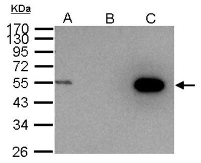
- Immunoprecipitation: Cyclin A2 Antibody [NBP1-31330] - Sample: 1000 ug 293T whole cell lysate/extract A. 30 ug 293T whole cell lysate/extract, B. Control with 2. 5 ug of preimmune rabbit IgG, C. Immunoprecipitation of Cyclin A2 protein by 2. 5 ug of Cyclin A2 antibody 10% SDS-PAGE gel.
Supportive validation
- Submitted by
- Novus Biologicals (provider)
- Main image
- Experimental details
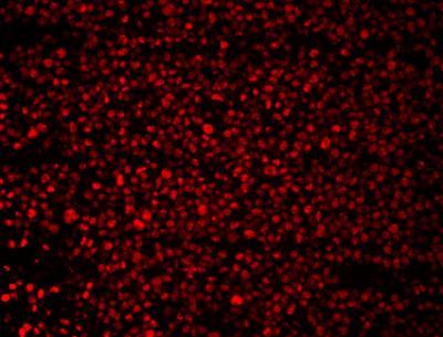
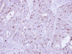
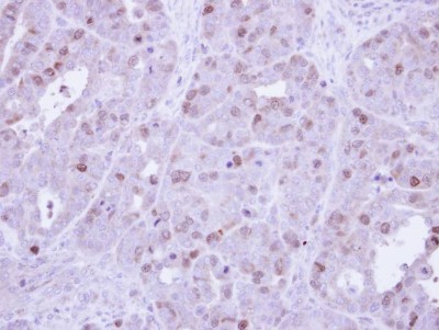
- Immunohistochemistry-Paraffin: Cyclin A2 Antibody [NBP1-31330] - Paraffin-embedded NCIN87 Xenograft, using antibody at 1:500 dilution.
- Submitted by
- Novus Biologicals (provider)
- Main image
- Experimental details
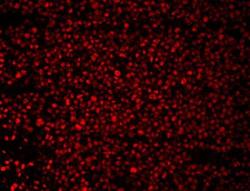
- Immunohistochemistry-Paraffin: Cyclin A2 Antibody [NBP1-31330] - 293T cells stained with Cyclin A2 antibody. Image from verified customer review.