Explore
 Validate
 Learn
LearnPA5-29767
antibody from Invitrogen Antibodies
Targeting: TAGLN
DKFZp686P11128, SM22, SMCC, TAGLN1, WS3-10
 Western blot
Antibody data
- Antibody Data
- Antigen structure
- References [2]
- Comments [0]
- Validations
- Western blot [5]
- Immunocytochemistry [1]
- Immunohistochemistry [3]
- Other assay [3]
Submit
Validation data
Reference
Comment
Report error
- Product number
- PA5-29767 - Provider product page

- Provider
- Invitrogen Antibodies
- Product name
- TAGLN Polyclonal Antibody
- Antibody type
- Polyclonal
- Antigen
- Recombinant protein fragment
- Description
- Recommended positive controls: 293T, A431, HeLa, HepG2, Mouse liver, A-10, A-10 treated with MG132, Bovine Smooth Muscle Cell, Bovine Fibroblastic Chondrocyte.
- Concentration
- 0.33 mg/mL
Submitted references Transgelin is a poor prognostic factor associated with advanced colorectal cancer (CRC) stage promoting tumor growth and migration in a TGFβ-dependent manner.
Transgelin is a TGFβ-inducible gene that regulates osteoblastic and adipogenic differentiation of human skeletal stem cells through actin cytoskeleston organization.
Elsafadi M, Manikandan M, Almalki S, Mahmood A, Shinwari T, Vishnubalaji R, Mobarak M, Alfayez M, Aldahmash A, Kassem M, Alajez NM
Cell death & disease 2020 May 11;11(5):341
Cell death & disease 2020 May 11;11(5):341
Transgelin is a TGFβ-inducible gene that regulates osteoblastic and adipogenic differentiation of human skeletal stem cells through actin cytoskeleston organization.
Elsafadi M, Manikandan M, Dawud RA, Alajez NM, Hamam R, Alfayez M, Kassem M, Aldahmash A, Mahmood A
Cell death & disease 2016 Aug 4;7(8):e2321
Cell death & disease 2016 Aug 4;7(8):e2321
No comments: Submit comment
Supportive validation
- Submitted by
- Invitrogen Antibodies (provider)
- Main image

- Experimental details
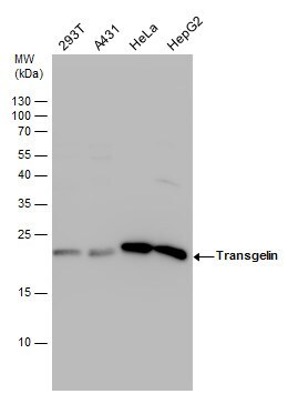
- Western blot analysis of Transgelin using Various whole cell extracts (30 µg). Samples were loaded onto a 15% SDS-PAGE gel and probed with a Transgelin polyclonal antibody (Product # PA5-29767) at a dilution of 1:1000.
- Submitted by
- Invitrogen Antibodies (provider)
- Main image
- Experimental details
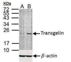
- Western Blot analysis of TAGLN was performed by separating 35 µg of whole cell lysates. Proteins were transferred to a membrane and probed with a TAGLN Polyclonal Antibody (Product # PA5-29767) at a dilution of 1:5000 (upper panel) and a beta-actin antibody (lower panel). The HRP-conjugated anti-rabbit IgG antibody was used to detect the primary antibody. A: A-10 cell , B. A-10 cell treated with MG132 .
- Submitted by
- Invitrogen Antibodies (provider)
- Main image

- Experimental details
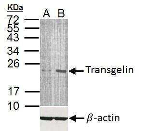
- Western Blot analysis of TAGLN was performed by separating 30 µg of various whole cell extracts by 15% SDS-PAGE. Proteins were transferred to a membrane and probed with a TAGLN Polyclonal Antibody (Product # PA5-29767) at a dilution of 1:1000 and a HRP-conjugated anti-rabbit IgG secondary antibody.
- Submitted by
- Invitrogen Antibodies (provider)
- Main image
- Experimental details
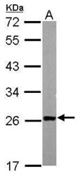
- Western Blot using TAGLN Polyclonal Antibody (Product # PA5-29767). Sample (50 µg of whole cell lysate). Lane A: mouse liver. 12% SDS PAGE. TAGLN Polyclonal Antibody (Product # PA5-29767) diluted at 1:1,000. The HRP-conjugated anti-rabbit IgG antibody was used to detect the primary antibody.

- Submitted by
- Invitrogen Antibodies (provider)
- Main image
- Experimental details
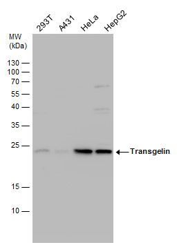
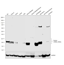
- Western blot was performed using Anti-TAGLN Polyclonal Antibody (Product # PA5-29767) and 23 and 20kDa bands corresponding to TAGLN were observed across all cell lines and tissues tested except Jurkat, Raji, Mouse Skeletal Muscle, Rat Skeletal Muscle and Rat Brain which are known to be low expressing (DOI: 10.1006/geno.1998.5267, 10.1371/journal.pone.0156429). Whole cell extracts (50 µg lysate) of BJ (Lane 1), Caco-2 (Lane 2), HeLa (Lane 3), Jurkat (Lane 4), Raji (Lane 5), Mouse Urinary Bladder (Lane 6), Mouse Skeletal Muscle (Lane 7), Rat Urinary Bladder (Lane 8), Rat Uterus (Lane 9), Rat Skeletal Muscle (Lane 10), Rat Brain (Lane 11) were electrophoresed using NuPAGE™ 4-12% Bis-Tris Protein Gel (Product # NP0322BOX). Resolved proteins were then transferred onto a Nitrocellulose membrane (Product # IB23001) by iBlot® 2 Dry Blotting System (Product # IB21001). The blot was probed with the primary antibody (1:1000 dilution) and detected by chemiluminescence with Goat anti-Rabbit IgG (H+L) Superclonal™ Secondary Antibody, HRP (Product # A27036, 1:4000 dilution) using the iBright FL 1000 (Product # A32752). Detection was performed using Novex® ECL Reagent Kit (Product # WP20005). TAGLN was observed in BJ, Caco-2, HeLa, Mouse Urinary Bladder, Rat Urinary Bladder and Rat Uterus but not in Jurkat, Raji, Mouse Skeletal Muscle, Rat Skeletal Muscle and Rat Brain which are known to be low expressing.

Supportive validation
- Submitted by
- Invitrogen Antibodies (provider)
- Main image

- Experimental details
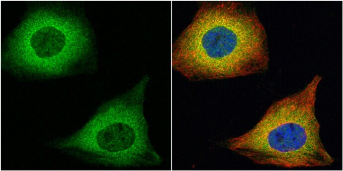
- Immunocytochemistry-Immunofluorescence analysis of TAGLN was performed in HeLa cells fixed in 4% paraformaldehyde at RT for 15 min. Green: TAGLN Polyclonal Antibody (Product # PA5-29767) diluted at 1:100. Red: alpha Tubulin, a cytoskeleton marker. Blue: Hoechst 33342 staining.
Supportive validation
- Submitted by
- Invitrogen Antibodies (provider)
- Main image
- Experimental details
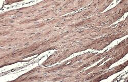
- TAGLN Polyclonal Antibody detects Transgelin protein at cytoplasm by immunohistochemical analysis. Sample: Paraffin-embedded mouse smooth muscle. Transgelin stained by TAGLN Polyclonal Antibody (Product # PA5-29767) diluted at 1:500. Antigen Retrieval: Citrate buffer, pH 6.0, 15 min.

- Submitted by
- Invitrogen Antibodies (provider)
- Main image
- Experimental details
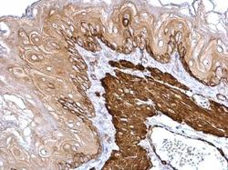
- TAGLN Polyclonal Antibody detects Transgelin protein at cytosol on mouse esophagus by immunohistochemical analysis. Sample: Paraffin-embedded mouse esophagus. TAGLN Polyclonal Antibody (Product # PA5-29767) dilution: 1:500. Antigen Retrieval: EDTA based buffer, pH 8.0, 15 min.

- Submitted by
- Invitrogen Antibodies (provider)
- Main image
- Experimental details
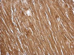
- TAGLN Polyclonal Antibody detects Transgelin protein at cytosol on mouse heart by immunohistochemical analysis. Sample: Paraffin-embedded mouse heart. TAGLN Polyclonal Antibody (Product # PA5-29767) dilution: 1:500. Antigen Retrieval: EDTA based buffer, pH 8.0, 15 min.

Supportive validation
- Submitted by
- Invitrogen Antibodies (provider)
- Main image
- Experimental details
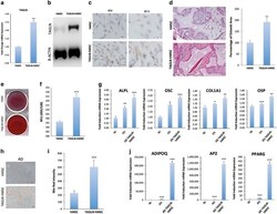
- Figure 3 TAGLN overexpression induces osteogenesis and adipogenesis in hMSC. ( a ) All controls represent empty vector-transfected cells. qRT-PCR of TAGLN gene expression in control (empty vector) hMSC and TAGLN-overexpressing line (TAGLN-hMSC). ( b ) Western blotting for TAGLN in TAGLN-hMSC (upper panel) and B-Actin (lower panel). ( c ) TAGLN immunocytochemical staining in control hMSC and TAGLN-hMSC cells at magnification x 40 and x 60. Cells were differentiated into osteoblasts by osteogenic or adipogenic induction mixture for 14 days. ( d ) hMSC and TAGLN-hMSC cells were implanted with HA/TCP subcutaneously into NOD/SCID mice. The histology of in vivo formed bone was examined with H&E staining (original magnification x 200). High-resolution whole-slide digital scans of all histological sections were created with a ScanScope scanner and bone formed was quantified. ( e ) Mineralized bone matrix formation in control hMSC (empty vector) cells and TAGLN-hMSC stained for Alizarin Red. ( f ) Quantification of Alizarin Red staining in control hMSC and TAGLN-hMSC. ( g ) qRT-PCR for osteogenic markers: ALPL, OSC, COL1A1 (collagen type I), and OSP: NI, OS, and osteoblast-induced TAGLN-hMSC (OS TAGLN-hMSC). ( h ) Both control (empty vector) hMSC, and TAGLN-hMSC were induced to adipocyte differentiation using adipocyte induction medium for 7 days. Mature lipid-filled adipocytes were stained by oil red. ( i ) Quantification of Nile Red staining for mature adipocytes: control (empty vec

- Submitted by
- Invitrogen Antibodies (provider)
- Main image

- Experimental details
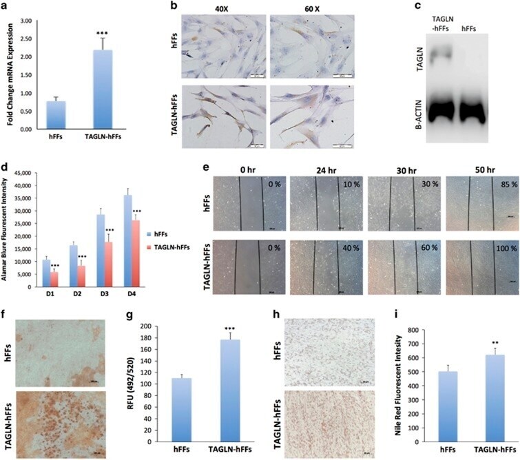
- Figure 4 Effects of TAGLN overexpression on hFFs functions. ( a ) qRT-PCR of TAGLN gene expression in control (empty vector) TAGLN-hFFs. ( b ) Immunocytochemical staining for TAGLN protein in control hFFs, and TAGLN-hFFs at magnifications x 40 and x 60. ( c ) Western blot of TAGLN (upper panel) and B-Actin (lower panel) in control hFFs and TAGLN-hFFs. Cells were induced to differentiate into osteoblasts or adipocytes by either osteogenic or adipogenic mixture for 24 days. ( d ) Alamar blue staining for cell viability in control hFFs and TAGLN-hFFs on day (D): 1D, 2D, 3D, and 4D. ( e ) Scratch assay where the motility of hFFs and TAGLN-hFFs cells was observed at different time intervals: 0, 24, 30, and 50 h. ( f ) Alizarin Red S staining for mineralized matrix formation. ( g ) Quantification of mineralized matrix formation in control hFFs cells and TAGLN-hFFs using Osteo-Image Mineralization Assay. ( h ) Control hFFs and hFF-TAGLN were induced to adipocyte differentiation by incubation in adipocyte induction medium for 24 days and stained by oil red for lipid-filled mature adipocytes. ( i ) Quantification of Nile red staining of mature adipocytes formed in control hFF cells and TAGLN-hFFs. Data are shown as mean+-S.D. of three independent experiments. ** P
- Submitted by
- Invitrogen Antibodies (provider)
- Main image

- Experimental details
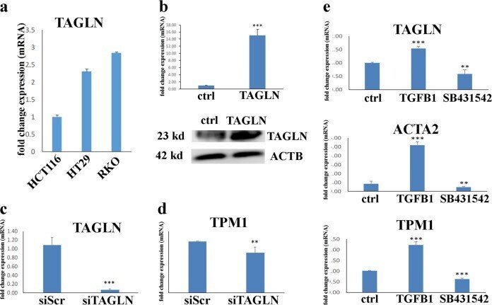
- Fig. 2 TAGLN is a TGFbeta responsive gene in CRC. a TAGLN mRNA expression in three different CRC cell lines based on the CCLE database: HCT116, HT-29, and RKO. Expression level is compared to TAGLN expression in HCT116 cells. b qRT-PCR of TAGLN expression in HCT116 control cells or cells transduced with TAGLN-overexpressing lentiviral vector (upper panel). Western blotting for TAGLN (upper panel) in HCT116-TAGLN vs. control cells. beta-Actin was used as the loading control. QRT-PCR for TAGLN ( c ) or TPM1 ( d ) gene expression 3 days post transfection of RKO cells with TAGLN-siRNA (siTAGLN) or scrambled-siRNA (siScr). Data are presented as fold change in mRNA expresion. e qRT-PCR of TAGLN, TPM1, and ACTA2 gene expression in RKO cells treated with TGFbeta1 (10 ng/ul) or SB431542 (10 µM), compared to control cells. The two-tailed t -test was used to compare different treatment groups. ** P < 0.005, *** P < 0.0005.