 Explore
 Validate
 Learn
Learn Western blot
 Immunocytochemistry
Antibody data
- Antibody Data
- Antigen structure
- References [0]
- Comments [0]
- Validations
- Western blot [1]
- Immunoprecipitation [1]
- Immunohistochemistry [2]
- Flow cytometry [1]
Submit
Validation data
Reference
Comment
Report error
- Product number
- NBP2-29905 - Provider product page

- Provider
- Novus Biologicals
- Product name
- Rabbit Polyclonal Pax5/BSAP Antibody
- Antibody type
- Polyclonal
- Description
- Protein G purified.
- Reactivity
- Human, Mouse
- Host
- Rabbit
- Isotype
- IgG
- Vial size
- 100 ug
- Concentration
- 1 mg/ml
- Storage
- Store at -20C. Avoid freeze-thaw cycles.
No comments: Submit comment
Supportive validation
- Submitted by
- Novus Biologicals (provider)
- Main image

- Experimental details
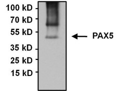
- Western Blot: Pax5/BSAP Antibody [NBP2-29905] - Analysis of 75ug of whole cell lysates from PAX5-expressing B cell lymphoma-derived cell lines, negative control Jurkat cells, and 10ul of PageRuler Plus Prestained Protein Ladder.

Supportive validation
- Submitted by
- Novus Biologicals (provider)
- Main image
- Experimental details
- Immunoprecipitation: Pax5/BSAP Antibody [NBP2-29905] - Analysis of PAX5 was performed using Raji whole cell lysates. Antigen-antibody complexes were formed by incubating 750ug of lysate with 5ug of a PAX5 polyclonal antibody overnight on a rocking platform at 4C. The immune complexes were captured on 50ul Protein A/G Agarose, washed extensively, and eluted with 5X Lane Marker Reducing Sample Buffer. Samples was resolved on a 4-20% Tris-HCl polyacrylamide gel, transferred to a PVDF membrane, and blocked with 5% BSA/TBS-0.1%Tween-20 for 1 hour. The membrane was probed with a PAX5 polyclonal antibody at a dilution of 1:1000 overnight rotating at 4C. Membranes were washed in TBST, and probed with Clean-Blot IP Detection Reagent at a dilution of 1:1000 for at least one hour. Chemiluminescent detection was performed using SuperSignal West Pico.

Supportive validation
- Submitted by
- Novus Biologicals (provider)
- Main image
- Experimental details
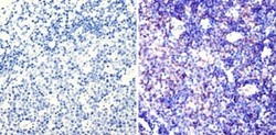
- Immunohistochemistry-Paraffin: Pax5/BSAP Antibody [NBP2-29905] - Analysis showing staining in the nucleus of mouse lymph node tissue (right) compared to a negative control without primary antibody (left).

- Submitted by
- Novus Biologicals (provider)
- Main image

- Experimental details
- Immunohistochemistry-Paraffin: Pax5/BSAP Antibody [NBP2-29905] - Analysis showing staining in the nucleus of human tonsil tissue (right) compared to a negative control without primary antibody (left).

Supportive validation
- Submitted by
- Novus Biologicals (provider)
- Main image

- Experimental details
- Flow Cytometry: Pax5/BSAP Antibody [NBP2-29905] - Analysis of PAX5 (red histogram) on Ramos cells. Cells were harvested, fixed with 4% formaldehyde, permeabilized, washed with PBS, and incubated with a PAX5 polyclonal antibody at a 1:50 dilution or with PBS alone (black histogram) for 1 hour at room temperature. For flow cytometry analysis, 30-minute incubation with DyLight 488 goat anti-rabbit IgG secondary antibody was performed. 30,000 cells were acquired for analysis.
