14-9918-82
antibody from Invitrogen Antibodies
Targeting: PAX5
BSAP
 Western blot
 Immunocytochemistry
 Immunoprecipitation
 Immunohistochemistry
 Chromatin Immunoprecipitation
 Other assay
Antibody data
- Antibody Data
- Antigen structure
- References [5]
- Comments [0]
- Validations
- Immunocytochemistry [2]
- Immunohistochemistry [1]
- Chromatin Immunoprecipitation [4]
- Other assay [4]
Submit
Validation data
Reference
Comment
Report error
- Product number
- 14-9918-82 - Provider product page

- Provider
- Invitrogen Antibodies
- Product name
- PAX5 Monoclonal Antibody (1H9), eBioscience™
- Antibody type
- Monoclonal
- Antigen
- Other
- Description
- Description: The monoclonal antibody 1H9 recognizes both mouse and human Pax5. Pax5, also known as BSAP (B cell specific activator protein), is a member of the paired box (pax) family of transcription factors. Pax5 is the only member of the pax family of transcription factors that is expressed in hematopoietic cells. During embryogenesis, Pax5 is transiently expressed in the brain of mice and in the mesencephalon and spinal cord of humans. Its expression is upregulated early in B cell development at the time of B cell commitment and is maintained throughout most subsequent stages. Suppression of Pax5 is essential for expression of Blimp-1 and the terminal differentiation of plasma cells. In the spleen, expression of Pax5 is higher in marginal zone B cells (B220+ CD21high CD23low) than in other B cells, especially the transition 1 stage (B220+ CD21- CD23-). In addition to its role in B cell development, Pax5 also affects VH-DJH heavy chain recombination as well as influencing the expression of many other B and non-B cell related proteins. Pax5 expression is correlated with many neoplasms. In diffuse large B cell lymphomas (DLBCL) and non-Hodgkin lymphomas Pax5 is often mutated while in B-cell ALL, expression levels are high. Additionally, translocation with elastin, IGH, ETV6, FOXP1, and EVI3 have been identified. eBioscience recommends that the Foxp3 Buffers (Product # 00-5521) be used for optimal results when using this antibody for intracellular staining and flow cytometric analysis. Applications Reported: This 1H9 antibody has been reported for use in immunoprecipitation, immunoblotting (WB), and immunohistology staining of frozen tissue sections. Applications Tested: This 1H9 antibody has been tested by western blot analysis. This can be used at less than or equal to 5 µg/mL. It is recommended that the antibody be carefully titrated for optimal performance in the assay of interest. Purity: Greater than 90%, as determined by SDS-PAGE. Aggregation: Less than 10%, as determined by HPLC. Filtration: 0.2 µm post-manufacturing filtered.
- Reactivity
- Human, Mouse
- Host
- Rat
- Isotype
- IgG
- Antibody clone number
- 1H9
- Vial size
- 100 μg
- Concentration
- 0.5 mg/mL
- Storage
- 4°C
Submitted references Asymmetric PI3K Activity in Lymphocytes Organized by a PI3K-Mediated Polarity Pathway.
The histone methyltransferase SETDB1 represses endogenous and exogenous retroviruses in B lymphocytes.
PDK1 regulates VDJ recombination, cell-cycle exit and survival during B-cell development.
Pax5: the guardian of B cell identity and function.
Reporter gene insertions reveal a strictly B lymphoid-specific expression pattern of Pax5 in support of its B cell identity function.
Chen YH, Kratchmarov R, Lin WW, Rothman NJ, Yen B, Adams WC, Nish SA, Rathmell JC, Reiner SL
Cell reports 2018 Jan 23;22(4):860-868
Cell reports 2018 Jan 23;22(4):860-868
The histone methyltransferase SETDB1 represses endogenous and exogenous retroviruses in B lymphocytes.
Collins PL, Kyle KE, Egawa T, Shinkai Y, Oltz EM
Proceedings of the National Academy of Sciences of the United States of America 2015 Jul 7;112(27):8367-72
Proceedings of the National Academy of Sciences of the United States of America 2015 Jul 7;112(27):8367-72
PDK1 regulates VDJ recombination, cell-cycle exit and survival during B-cell development.
Venigalla RK, McGuire VA, Clarke R, Patterson-Kane JC, Najafov A, Toth R, McCarthy PC, Simeons F, Stojanovski L, Arthur JS
The EMBO journal 2013 Apr 3;32(7):1008-22
The EMBO journal 2013 Apr 3;32(7):1008-22
Pax5: the guardian of B cell identity and function.
Cobaleda C, Schebesta A, Delogu A, Busslinger M
Nature immunology 2007 May;8(5):463-70
Nature immunology 2007 May;8(5):463-70
Reporter gene insertions reveal a strictly B lymphoid-specific expression pattern of Pax5 in support of its B cell identity function.
Fuxa M, Busslinger M
Journal of immunology (Baltimore, Md. : 1950) 2007 Mar 1;178(5):3031-7
Journal of immunology (Baltimore, Md. : 1950) 2007 Mar 1;178(5):3031-7
No comments: Submit comment
Supportive validation
- Submitted by
- Invitrogen Antibodies (provider)
- Main image
- Experimental details
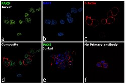
- Immunofluorescence analysis of PAX5 was performed using 70% confluent log phase Ramos cells. The cells were fixed with 4% Paraformaldehyde for 10 minutes, permeabilized with 0.1% Triton™ X-100 for 10 minutes, and blocked with 2% BSA for 10 minutes at room temperature. The cells were labeled with PAX5 Monoclonal Antibody (1H9), eBioscience™ (Product # 14-9918-82) at 5 µg/mL in 0.1% BSA, incubated at 4 degree Celsius overnight and then labeled with Goat anti-Rat IgG (H+L) Cross-Adsorbed Secondary Antibody, Alexa Fluor 488 (Product # A-11006), (1:2000 dilution) for 45 minutes at room temperature (Panel a: Green). Nuclei (Panel b: Blue) were stained with SlowFade® Gold Antifade Mountant with DAPI (Product # S36938). F-actin (Panel c: Red) was stained with Rhodamine Phalloidin (Product # R415, 1:300). Panel d represents the merged image showing nuclear localization. Panel e represents Jurkat cells having no expression of PAX5. Panel f represents control cells with no primary antibody to assess background. The images were captured at 60X magnification.
- Submitted by
- Invitrogen Antibodies (provider)
- Main image
- Experimental details
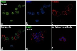
- Immunofluorescence analysis of PAX5 was performed using 70% confluent log phase Ramos cells. The cells were fixed with 4% Paraformaldehyde for 10 minutes, permeabilized with 0.1% Triton™ X-100 for 10 minutes, and blocked with 2% BSA for 10 minutes at room temperature. The cells were labeled with PAX5 Monoclonal Antibody (1H9), eBioscience™ (Product # 14-9918-82) at 5 µg/mL in 0.1% BSA, incubated at 4 degree Celsius overnight and then labeled with Goat anti-Rat IgG (H+L) Cross-Adsorbed Secondary Antibody, Alexa Fluor 488 (Product # A-11006), (1:2000 dilution) for 45 minutes at room temperature (Panel a: Green). Nuclei (Panel b: Blue) were stained with SlowFade® Gold Antifade Mountant with DAPI (Product # S36938). F-actin (Panel c: Red) was stained with Rhodamine Phalloidin (Product # R415, 1:300). Panel d represents the merged image showing nuclear localization. Panel e represents Jurkat cells having no expression of PAX5. Panel f represents control cells with no primary antibody to assess background. The images were captured at 60X magnification.
Supportive validation
- Submitted by
- Invitrogen Antibodies (provider)
- Main image
- Experimental details
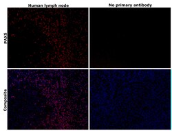
- Immunohistochemical analysis of PAX5 was performed using formalin-fixed paraffin-embedded human lymph node tissue sections. To expose the target protein, heat-induced epitope retrieval was performed on de-paraffinized sections using eBioscience™ IHC Antigen Retrieval Solution - High pH (10X) (Product # 00-4956-58) diluted to 1X solution in water in a decloaking chamber at 110 degree Celsius for 15 minutes. Following antigen retrieval, the sections were blocked with 2% normal goat serum in 1X PBS for 45 minutes at room temperature and then probed with or without PAX5 Monoclonal Antibody (1H9), eBioscience™ (Product # 14-9918-82) at 5 µg/mL in 0.1% normal goat serum overnight at 4 degree Celsius in a humidified chamber. Detection was performed using Goat anti-Rat IgG (H+L) Cross-Adsorbed Secondary Antibody, Alexa Fluor™ 647 (Product# A-21247) at a dilution of 1:2,000 dilution in 0.1% normal goat serum for 45 minutes at room temperature. ReadyProbes™ Tissue Autofluorescence Quenching Kit (Product # R37630) was used to quench autofluorescence from the tissues. Nuclei were stained with DAPI (Product # D1306) and the sections were mounted using ProLong™ Glass Antifade Mountant (Product # P36984). The images were captured on EVOS™ M7000 Imaging System (Product # AMF7000) at 20X magnification and externally deconvoluted.
Supportive validation
- Submitted by
- Invitrogen Antibodies (provider)
- Main image
- Experimental details
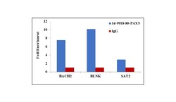
- Chromatin Immunoprecipitation (ChIP) assay of endogenous PAX5 protein using Anti-PAX5 Antibody: ChIP was performed using Anti-PAX5 Monoclonal Antibody (Product # 14-9918-80, 5 µg) on sheared chromatin from Raji cells using the MAGnify ChIP System kit (Product # 49-2024). Normal Mouse IgG was used as a negative IP control. The purified DNA was analyzed by qPCR using primers binding to BACH2 and BLNK promoter and SAT2 satellite repeats. Data is presented as fold enrichment of the antibody signal versus the negative control IgG using the comparative CT method.
- Submitted by
- Invitrogen Antibodies (provider)
- Main image

- Experimental details
- Chromatin Immunoprecipitation (ChIP) assay of endogenous PAX5 protein using Anti-PAX5 Antibody: ChIP was performed using Anti-PAX5 Monoclonal Antibody (Product # 14-9918-82, 5 µg) on sheared chromatin from Raji cells using the MAGnify ChIP System kit (Product # 49-2024). Normal Mouse IgG was used as a negative IP control. The purified DNA was analyzed by qPCR using primers binding to BACH2 and BLNK promoter and SAT2 satellite repeats. Data is presented as fold enrichment of the antibody signal versus the negative control IgG using the comparative CT method.
- Submitted by
- Invitrogen Antibodies (provider)
- Main image
- Experimental details
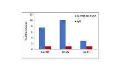
- Chromatin Immunoprecipitation (ChIP) assay of endogenous PAX5 protein using Anti-PAX5 Antibody: ChIP was performed using Anti-PAX5 Monoclonal Antibody (Product # 14-9918-80, 5 µg) on sheared chromatin from Raji cells using the MAGnify ChIP System kit (Product # 49-2024). Normal Mouse IgG was used as a negative IP control. The purified DNA was analyzed by qPCR using primers binding to BACH2 and BLNK promoter and SAT2 satellite repeats. Data is presented as fold enrichment of the antibody signal versus the negative control IgG using the comparative CT method.
- Submitted by
- Invitrogen Antibodies (provider)
- Main image
- Experimental details
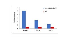
- Chromatin Immunoprecipitation (ChIP) assay of endogenous PAX5 protein using Anti-PAX5 Antibody: ChIP was performed using Anti-PAX5 Monoclonal Antibody (Product # 14-9918-82, 5 µg) on sheared chromatin from Raji cells using the MAGnify ChIP System kit (Product # 49-2024). Normal Mouse IgG was used as a negative IP control. The purified DNA was analyzed by qPCR using primers binding to BACH2 and BLNK promoter and SAT2 satellite repeats. Data is presented as fold enrichment of the antibody signal versus the negative control IgG using the comparative CT method.
Supportive validation
- Submitted by
- Invitrogen Antibodies (provider)
- Main image
- Experimental details
- NULL
- Submitted by
- Invitrogen Antibodies (provider)
- Main image

- Experimental details
- NULL
- Submitted by
- Invitrogen Antibodies (provider)
- Main image

- Experimental details
- Figure 7 PDK1 regulates Pax5 expression and cell-cycle progression in pre-B cells. To look at proliferation in vivo , PDK1 +/+ /Vav-Cre +ve and PDK1 fl/fl /Vav-Cre +ve mice were injected with 1 mg BrdU 3.25 h before being sacrificed. Bone marrow was then stained for 7-AAD and pre-B cell markers as described in Materials and methods. BrdU and 7-AAD uptake was determined by FACS in the pro-B and small pre-B cells ( A ). The levels of the proliferation marker Ki67 as well as cyclinD3 in ex vivo pre-B cells were also determined by FACS ( B ). To examine the expression of Pax5, IRF4, IRF8, Ikaros and Aiolos, total RNA was isolated from FACS-sorted pre-B cells and the mRNA levels for these genes determined by qPCR ( C ). Error bars represent the standard deviation of RNA from FACS-sorted pre-B cells of three independent pools of mice per genotype, P 0.05. The levels of Pax5, IRF4, IRF8, Ikaros and Aiolos protein ( D ) were determined by intracellular FACS staining of pro-B (CD19 +ve IgM -ve CD43 high B220 +ve ), large pre-B (FSC high CD19 +ve IgM -ve CD43 -ve B220 +ve ) or small pre-B (FSC low CD19 +ve IgM -ve CD43 -ve B220 +ve ). Results are representative of three mice per genotype.
- Submitted by
- Invitrogen Antibodies (provider)
- Main image

- Experimental details
- Figure 8 Pax5 and Bcl2A1 induce B-cell differentiation in the absence of PDK1. The levels of apoptosis in ex vivo PDK1 +/+ Vav-Cre +ve and PDK1 fl/fl Vav-Cre +ve cells were determined by annexin V staining of pro-B (CD19 +ve IgM -ve CD43 high B220 +ve ), large pre-B (FSC high CD19 +ve IgM -ve CD43 -ve B220 +ve ) or small pre-B (FSC low CD19 +ve IgM -ve CD43 -ve B220 +ve ) cells. Results are representative of three mice ( A ). qPCR was used to determine the mRNA levels of Mcl-1 and Bcl2A1 in FACS-sorted pre-B cells ( n =3) from PDK1 +/+ Vav-Cre +ve and PDK1 fl/fl Vav-Cre +ve mice ( B ). To determine if Pax5 and Bcl2A1 were involved in mediating PDK1 function in B cells, PDK1 fl/fl Vav-Cre +ve FACS-sorted CD19 +ve IgM -ve CD43 high B220 +ve DAPI -ve pro-B cells were infected with the indicated combinations of retroviruses for GFP, mCherry, GFP-Pax5 or mCherry-Bcl2A1 as described in Materials and methods. The percentages of live cells on day 6 in culture were determined based on forward and side scatter ( C , upper panels). BCR expression was then determined by surface staining for IgM and IgD in the GFP and/or mCherry-positive cells ( C , lower panels). Quantification of the % of live cells in ( C ) ( n =3) is shown in ( D ) while qualification of the % IgM/IgD positive gates for the indicated proteins in shown in ( E ). To analyse the effect on the cell cycle, PDK1 knockout pro-B cells were isolated and transfected with retroviruses for GFP-Pax5 and mCherry-Bcl2A1. After 6 day
 Explore
 Validate
 Learn
Learn