 Explore
 Validate
 Learn
Learn Western blot
 Immunocytochemistry
Antibody data
- Antibody Data
- Antigen structure
- References [0]
- Comments [0]
- Validations
- Western blot [2]
- Immunohistochemistry [1]
Submit
Validation data
Reference
Comment
Report error
- Product number
- NB100-1768 - Provider product page

- Provider
- Novus Biologicals
- Proper citation
- Novus Cat#NB100-1768, RRID:AB_10003514
- Product name
- Mouse Monoclonal Chk1 Antibody
- Antibody type
- Monoclonal
- Description
- Protein G purified.
- Reactivity
- Human
- Host
- Mouse
- Isotype
- IgG
- Vial size
- 0.1 ml
- Concentration
- 1.0 mg/ml
- Storage
- Store at 4C short term. Aliquot and store at -20C long term. Avoid freeze-thaw cycles.
No comments: Submit comment
Supportive validation
- Submitted by
- Novus Biologicals (provider)
- Main image

- Experimental details
- Western Blot: Chk1 Antibody (2G1D5) [NB100-1768] - Analysis using anti-Chk1 mouse mAb against A431 (1), Hela (2), NIH/3T3 (3) and K562 (4) cell lysates.

- Submitted by
- Novus Biologicals (provider)
- Main image
- Experimental details
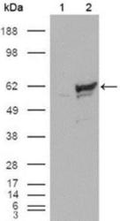
- Western Blot: Chk1 Antibody (2G1D5) [NB100-1768] - Analysis using Chk1 mouse mAb against HEK293T cells transfected with the pCMV6-ENTRY control (1) and pCMV6-ENTRY Chk1 cDNA (2).

Supportive validation
- Submitted by
- Novus Biologicals (provider)
- Main image

- Experimental details
- Immunohistochemistry-Paraffin: Chk1 Antibody (2G1D5) [NB100-1768] - Analysis of FFPE human lung using Chk1 antibody (clone 2G1D5) at 1:250 on a Bond Rx autostainer (Leica Biosystems). The assay involved 20 minutes of heat induced antigen retrieval (HIER) using 10 mM sodium citrate buffer (pH 6.0) and endogenous peroxidase quenching with peroxide block. The sections were incubated with primary antibody for 30 minutes and Bond Polymer Refine Detection (Leica Biosystems) with DAB was used for signal development followed by counterstaining with hematoxylin. Whole slide scanning and capturing of representative images was performed using Aperio AT2 (Leica Biosystems). Cytoplasmic and nuclear staining of Chk1 was observed as this is a ubiquitous cell checkpoint protein. Staining was performed by Histowiz.
