 Explore
 Validate
 Learn
Learn Western blot
 Flow cytometry
Antibody data
- Antibody Data
- Antigen structure
- References [16]
- Comments [0]
- Validations
- Western blot [1]
- Immunocytochemistry [4]
- Immunoprecipitation [1]
- Immunohistochemistry [3]
Submit
Validation data
Reference
Comment
Report error
- Product number
- SM5072 - Provider product page

- Provider
- Acris Antibodies GmbH
- Proper citation
- Acris Antibodies GmbH Cat#SM5072, RRID:AB_1002596
- Product name
- anti HSPA8 / HSC70
- Antibody type
- Monoclonal
- Antigen
- Mouse spermatogenic cell protein.
- Reactivity
- Human, Mouse, Rat, Bovine, Feline, Hamster
- Host
- Mouse
- Isotype
- IgM
- Antibody clone number
- 13D3
- Vial size
- 50 µl
- Concentration
- 5.6 mg/ml
Submitted references Loss of nuclear pro-IL-16 facilitates cell cycle progression in human cutaneous T cell lymphoma.
A sporadic Parkinson disease model via silencing of the ubiquitin-proteasome/E3 ligase component SKP1A.
Heat shock enhances the susceptibility of BHK cells to rotavirus infection through the facilitation of entry and post-entry virus replication steps.
Beta-amyloid treatment of two complementary P301L tau-expressing Alzheimer's disease models reveals similar deregulated cellular processes.
Proteomic analysis of exosomes secreted by human mesothelioma cells.
The repeated bout effect and heat shock proteins: intramuscular HSP27 and HSP70 expression following two bouts of eccentric exercise in humans.
Heat shock cognate protein 70 is involved in rotavirus cell entry.
Characterization and regulation of the major histocompatibility complex-encoded proteins Hsp70-Hom and Hsp70-1/2.
Heat shock cognate-70 gene expression declines during normal aging of the primate retina.
Blocking the endogenous increase in HSP 72 increases susceptibility to hypoxia and reoxygenation in isolated adult feline cardiocytes.
A 16-kDa protein functions as a new regulatory protein for Hsc70 molecular chaperone and is identified as a member of the Nm23/nucleoside diphosphate kinase family.
Platelet adhesion to collagen under flow causes dissociation of a phosphoprotein complex of heat-shock proteins and protein phosphatase 1.
Heat shock factor-1 and the heat shock cognate 70 protein associate in high molecular weight complexes in the cytoplasm of NIH-3T3 cells.
Protein and peptide binding and stimulation of in vitro lysosomal proteolysis by the 73-kDa heat shock cognate protein.
Heat-shock cognate protein (hsc71) and related proteins in mouse spermatogenic cells.
A novel hsp70-like protein (P70) is present in mouse spermatogenic cells.
Curiel-Lewandrowski C, Yamasaki H, Si CP, Jin X, Zhang Y, Richmond J, Tuzova M, Wilson K, Sullivan B, Jones D, Ryzhenko N, Little F, Kupper TS, Center DM, Cruikshank WW
The Journal of clinical investigation 2011 Dec;121(12):4838-49
The Journal of clinical investigation 2011 Dec;121(12):4838-49
A sporadic Parkinson disease model via silencing of the ubiquitin-proteasome/E3 ligase component SKP1A.
Fishman-Jacob T, Reznichenko L, Youdim MB, Mandel SA
The Journal of biological chemistry 2009 Nov 20;284(47):32835-45
The Journal of biological chemistry 2009 Nov 20;284(47):32835-45
Heat shock enhances the susceptibility of BHK cells to rotavirus infection through the facilitation of entry and post-entry virus replication steps.
López T, López S, Arias CF
Virus research 2006 Oct;121(1):74-83
Virus research 2006 Oct;121(1):74-83
Beta-amyloid treatment of two complementary P301L tau-expressing Alzheimer's disease models reveals similar deregulated cellular processes.
David DC, Ittner LM, Gehrig P, Nergenau D, Shepherd C, Halliday G, Götz J
Proteomics 2006 Dec;6(24):6566-77
Proteomics 2006 Dec;6(24):6566-77
Proteomic analysis of exosomes secreted by human mesothelioma cells.
Hegmans JP, Bard MP, Hemmes A, Luider TM, Kleijmeer MJ, Prins JB, Zitvogel L, Burgers SA, Hoogsteden HC, Lambrecht BN
The American journal of pathology 2004 May;164(5):1807-15
The American journal of pathology 2004 May;164(5):1807-15
The repeated bout effect and heat shock proteins: intramuscular HSP27 and HSP70 expression following two bouts of eccentric exercise in humans.
Thompson HS, Clarkson PM, Scordilis SP
Acta physiologica Scandinavica 2002 Jan;174(1):47-56
Acta physiologica Scandinavica 2002 Jan;174(1):47-56
Heat shock cognate protein 70 is involved in rotavirus cell entry.
Guerrero CA, Bouyssounade D, Zárate S, Isa P, López T, Espinosa R, Romero P, Méndez E, López S, Arias CF
Journal of virology 2002 Apr;76(8):4096-102
Journal of virology 2002 Apr;76(8):4096-102
Characterization and regulation of the major histocompatibility complex-encoded proteins Hsp70-Hom and Hsp70-1/2.
Fourie AM, Peterson PA, Yang Y
Cell stress & chaperones 2001 Jul;6(3):282-95
Cell stress & chaperones 2001 Jul;6(3):282-95
Heat shock cognate-70 gene expression declines during normal aging of the primate retina.
Bernstein SL, Liu AM, Hansen BC, Somiari RI
Investigative ophthalmology & visual science 2000 Sep;41(10):2857-62
Investigative ophthalmology & visual science 2000 Sep;41(10):2857-62
Blocking the endogenous increase in HSP 72 increases susceptibility to hypoxia and reoxygenation in isolated adult feline cardiocytes.
Nakano M, Mann DL, Knowlton AA
Circulation 1997 Mar 18;95(6):1523-31
Circulation 1997 Mar 18;95(6):1523-31
A 16-kDa protein functions as a new regulatory protein for Hsc70 molecular chaperone and is identified as a member of the Nm23/nucleoside diphosphate kinase family.
Leung SM, Hightower LE
The Journal of biological chemistry 1997 Jan 31;272(5):2607-14
The Journal of biological chemistry 1997 Jan 31;272(5):2607-14
Platelet adhesion to collagen under flow causes dissociation of a phosphoprotein complex of heat-shock proteins and protein phosphatase 1.
Polanowska-Grabowska R, Simon CG Jr, Falchetto R, Shabanowitz J, Hunt DF, Gear AR
Blood 1997 Aug 15;90(4):1516-26
Blood 1997 Aug 15;90(4):1516-26
Heat shock factor-1 and the heat shock cognate 70 protein associate in high molecular weight complexes in the cytoplasm of NIH-3T3 cells.
Nunes SL, Calderwood SK
Biochemical and biophysical research communications 1995 Aug 4;213(1):1-6
Biochemical and biophysical research communications 1995 Aug 4;213(1):1-6
Protein and peptide binding and stimulation of in vitro lysosomal proteolysis by the 73-kDa heat shock cognate protein.
Terlecky SR, Chiang HL, Olson TS, Dice JF
The Journal of biological chemistry 1992 May 5;267(13):9202-9
The Journal of biological chemistry 1992 May 5;267(13):9202-9
Heat-shock cognate protein (hsc71) and related proteins in mouse spermatogenic cells.
Maekawa M, O'Brien DA, Allen RL, Eddy EM
Biology of reproduction 1989 Apr;40(4):843-52
Biology of reproduction 1989 Apr;40(4):843-52
A novel hsp70-like protein (P70) is present in mouse spermatogenic cells.
Allen RL, O'Brien DA, Eddy EM
Molecular and cellular biology 1988 Feb;8(2):828-32
Molecular and cellular biology 1988 Feb;8(2):828-32
No comments: Submit comment
Supportive validation
- Submitted by
- Acris Antibodies GmbH (provider)
- Main image
- Experimental details
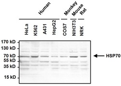
- Western blot analysis of Heat Shock Protein 70 (HSP70) was performed by loading 50µg of the indicated whole cell lysates and 15µl of PageRuler Prestained Protein Ladder onto a 4-20% Tris-HCl polyacrylamide gel. Proteins were transferred to a PVDF membrane and blocked with 5% BSA/TBST for at least 1 hour. The membrane was probed with a HSP70 monoclonal antibody (Cat.-No SM5072) at a dilution of 1:1000 overnight at 4°C on a rocking platform, washed in TBS-0.1%Tween 20, and probed with a goat anti-mouse IgM-HRP secondary antibody at a dilution of 1:20,000 for at least 1 hour. Chemiluminescent detection was performed using SuperSignal West Pico.

Supportive validation
- Submitted by
- Acris Antibodies GmbH (provider)
- Main image

- Experimental details
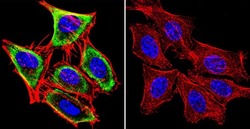
- Immunofluorescent analysis of Heat Shock Protein 70 using Heat Shock Protein 70 Monoclonal antibody (13D3) (Cat.-No SM5072) shows staining in MCF-7 cells. Heat Shock Protein 70 staining (green), F-Actin staining with Phalloidin (red) and nuclei with DAPI (blue) is shown. Cells were grown on chamber slides and fixed with formaldehyde prior to staining. Cells were probed without (control) or with or an antibody recognizing Heat Shock Protein 70 (Cat.-No SM5072) at a dilution of 1:20-1:200 over night at 4°C, washed with PBS and incubated with a DyLight-488 conjugated secondary antibody. Images were taken at 60X magnification.

- Submitted by
- Acris Antibodies GmbH (provider)
- Main image
- Experimental details
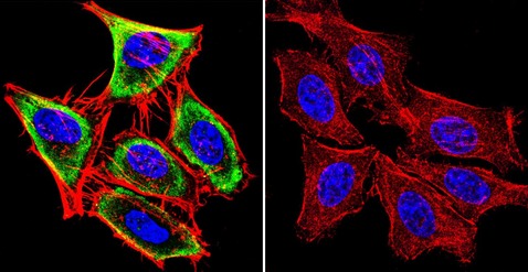
- Immunofluorescent analysis of Heat Shock Protein 70 using Heat Shock Protein 70 Monoclonal antibody (13D3) (Cat.-No SM5072) shows staining in HeLa cells. Heat Shock Protein 70 staining (green), F-Actin staining with Phalloidin (red) and nuclei with DAPI (blue) is shown. Cells were grown on chamber slides and fixed with formaldehyde prior to staining. Cells were probed without (control) or with or an antibody recognizing Heat Shock Protein 70 (Cat.-No SM5072) at a dilution of 1:20-1:200 over night at 4°C, washed with PBS and incubated with a DyLight-488 conjugated secondary antibody. Images were taken at 60X magnification.
- Submitted by
- Acris Antibodies GmbH (provider)
- Main image
- Experimental details
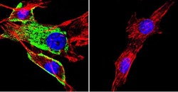
- Immunofluorescent analysis of Heat Shock Protein 70 using Heat Shock Protein 70 Monoclonal antibody (13D3) shows staining in NIH-3T3 cells. Heat Shock Protein 70 staining (green), F-Actin staining with Phalloidin (red) and nuclei with DAPI (blue) is shown. Cells were grown on chamber slides and fixed with formaldehyde prior to staining. Cells were probed without (control) or with or an antibody recognizing Heat Shock Protein 70 (Cat.-No SM5072) at a dilution of 1:20-1:200 over night at 4°C, washed with PBS and incubated with a DyLight-488 conjugated secondary antibody. Images were taken at 60X magnification.

- Submitted by
- Acris Antibodies GmbH (provider)
- Main image

- Experimental details
- Immunofluorescent analysis of Heat Shock Protein 70 (HSP70) (green) in HeLa and NIH3T3 cells. Formalin fixed cells were permeabilized with 0.1% Triton X-100 in TBS for 10 minutes at room temperature and blocked with 1% Blocker BSA for 15 minutes at room temperature. Cells were probed with a HSP70 monoclonal antibody (Cat.-No SM5072) at a dilution of 1:50 for at least 1 hour at room temperature, washed with PBS, and incubated with DyLight 488 goat anti-mouse IgG secondary antibody at a dilution of 1:400 for 30 minutes at room temperature. Nuclei (blue) were stained with Hoechst 33342 dye. Images were taken on a Thermo Scientific ArrayScan at 20X magnification.

Supportive validation
- Submitted by
- Acris Antibodies GmbH (provider)
- Main image
- Experimental details
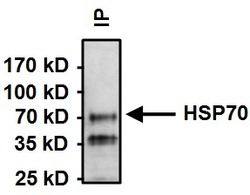
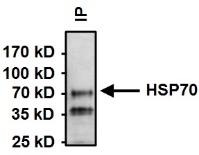
- Immunoprecipitation of Heat Shock Protein 70 (HSP70) was performed on HeLa cells. Antigen-antibody complexes were formed by incubating 500ug whole cell lysate with 2ul of HSP70 monoclonal antibody (Cat.-No SM5072) overnight on a rocking platform at 4°C. The immune complexes were captured on 50ul Protein A/G Plus Agarose, washed extensively, and eluted with Lane Marker Reducing Sample Buffer. Samples were resolved on a 4-20% Tris-HCl polyacrylamide gel, transferred to a PVDF membrane, and blocked with 5% BSA/TBST for at least 1 hour. The membrane was probed with a HSP70 monoclonal antibody (Cat.-No SM5072) at a dilution of 1:1000 overnight rotating at 4°C, washed in TBST, and probed with goat anti-mouse IgM-HRP secondary antibody for at least 1 hour. Chemiluminescent detection was performed using SuperSignal West Dura.
Supportive validation
- Submitted by
- Acris Antibodies GmbH (provider)
- Main image
- Experimental details
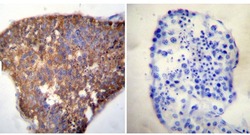
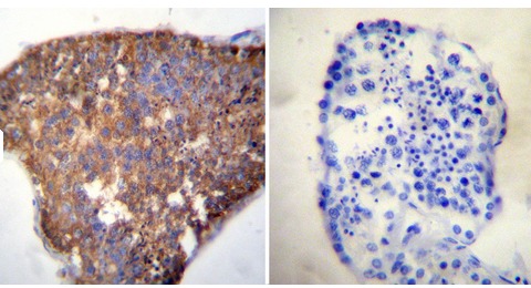
- Immunohistochemistry was performed on normal deparaffinized human Testis tissue. To expose target proteins, heat induced antigen retrieval was performed using 10mM sodium citrate (pH6.0) buffer, microwaved for 8-15 minutes. Following antigen retrieval tissues were blocked in 3% BSA-PBS for 30 minutes at room temperature. Tissues were then probed at a dilution of 1:20 with a mouse monoclonal antibody recognizing Heat Shock Cognate Protein 70 (Cat.-No SM5072) or without primary antibody (negative control) overnight at 4°C in a humidified chamber. Tissues were washed extensively with PBST and endogenous peroxidase activity was quenched with a peroxidase suppressor. Detection was performed using a biotin-conjugated secondary antibody and SA-HRP, followed by colorimetric detection using DAB. Tissues were counterstained with hematoxylin and prepped for mounting.
- Submitted by
- Acris Antibodies GmbH (provider)
- Main image

- Experimental details
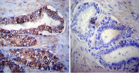
- Immunohistochemistry was performed on cancer biopsies of deparaffinized human Prostate carcinoma tissue. To expose target proteins, heat induced antigen retrieval was performed using 10mM sodium citrate (pH6.0) buffer, microwaved for 8-15 minutes. Following antigen retrieval tissues were blocked in 3% BSA-PBS for 30 minutes at room temperature. Tissues were then probed at a dilution of 1:200 with a mouse monoclonal antibody recognizing Heat Shock Cognate Protein 70 (Cat.-No SM5072) or without primary antibody (negative control) overnight at 4°C in a humidified chamber. Tissues were washed extensively with PBST and endogenous peroxidase activity was quenched with a peroxidase suppressor. Detection was performed using a biotin-conjugated secondary antibody and SA-HRP, followed by colorimetric detection using DAB. Tissues were counterstained with hematoxylin and prepped for mounting.
- Submitted by
- Acris Antibodies GmbH (provider)
- Main image

- Experimental details
- Immunohistochemistry was performed on normal deparaffinized human Tonsil tissue. To expose target proteins, heat induced antigen retrieval was performed using 10mM sodium citrate (pH6.0) buffer, microwaved for 8-15 minutes. Following antigen retrieval tissues were blocked in 3% BSA-PBS for 30 minutes at room temperature. Tissues were then probed at a dilution of 1:20 with a mouse monoclonal antibody recognizing Heat Shock Cognate Protein 70 (Cat.-No SM5072) or without primary antibody (negative control) overnight at 4°C in a humidified chamber. Tissues were washed extensively with PBST and endogenous peroxidase activity was quenched with a peroxidase suppressor. Detection was performed using a biotin-conjugated secondary antibody and SA-HRP, followed by colorimetric detection using DAB. Tissues were counterstained with hematoxylin and prepped for mounting.
