Explore
 Validate
 Learn
LearnNB100-1972
antibody from Novus Biologicals
Targeting: HSP90AA1
FLJ31884, Hsp89, Hsp90, HSP90N, HSPC1, HSPCA
 Western blot
 Immunocytochemistry
Antibody data
- Antibody Data
- Antigen structure
- References [3]
- Comments [0]
- Validations
- Western blot [1]
- Immunohistochemistry [1]
- Flow cytometry [6]
Submit
Validation data
Reference
Comment
Report error
- Product number
- NB100-1972 - Provider product page

- Provider
- Novus Biologicals
- Proper citation
- Novus Cat#NB100-1972, RRID:AB_2120909
- Product name
- Mouse Monoclonal HSP90 Antibody
- Antibody type
- Monoclonal
- Description
- Protein G purified. Recognizes both Hsp90 alpha and beta. It reacts with the rodent glucocorticoid receptor and with proliferation potential proteins (P2PS) with apparent molecular masses of 30-40 kDa.
- Reactivity
- Human, Mouse, Rat, Bacteria, Canine, Chicken/Avian, Guinea Pig, Porcine, Rabbit, Sheep, Simian
- Host
- Mouse
- Antigen sequence
The epitope of this antibody has be
en mapped to amino acid residues 60
4-697 of the human Hsp90 sequence.- Isotype
- IgG
- Vial size
- 0.1 mg
- Concentration
- 1.0 mg/ml
- Storage
- Store at 4C short term. Aliquot and store at -20C long term. Avoid freeze-thaw cycles.
Submitted references Medical Gas Plasma Jet Technology Targets Murine Melanoma in an Immunogenic Fashion.
Hmox1 Upregulation Is a Mutual Marker in Human Tumor Cells Exposed to Physical Plasma-Derived Oxidants.
Chaperone proteins as single component reagents to assess antibody nonspecificity.
Bekeschus S, Clemen R, Nießner F, Sagwal SK, Freund E, Schmidt A
Advanced science (Weinheim, Baden-Wurttemberg, Germany) 2020 May;7(10):1903438
Advanced science (Weinheim, Baden-Wurttemberg, Germany) 2020 May;7(10):1903438
Hmox1 Upregulation Is a Mutual Marker in Human Tumor Cells Exposed to Physical Plasma-Derived Oxidants.
Bekeschus S, Freund E, Wende K, Gandhirajan RK, Schmidt A
Antioxidants (Basel, Switzerland) 2018 Oct 27;7(11)
Antioxidants (Basel, Switzerland) 2018 Oct 27;7(11)
Chaperone proteins as single component reagents to assess antibody nonspecificity.
Kelly RL, Geoghegan JC, Feldman J, Jain T, Kauke M, Le D, Zhao J, Wittrup KD
mAbs 2017 Oct;9(7):1036-1040
mAbs 2017 Oct;9(7):1036-1040
No comments: Submit comment
Supportive validation
- Submitted by
- Novus Biologicals (provider)
- Main image
- Experimental details
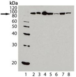
- Western Blot: HSP90 Antibody (AC88) [NB100-1972] - Analysis of HSP90, mAb (AC88): Lane 1: MW Marker; Lane 2: HSP90 (human), (native) ; Lane 3: HSP90 beta, (human) (recombinant); Lane 4: HSP90a (human), (recombinant); Lane 5: HeLa (heat shocked); Lane 6: 3T3 (heat shocked); Lane 7: PC-12 (heat shocked) ; Lane 8: CHO-K1 (heat shocked).
Supportive validation
- Submitted by
- Novus Biologicals (provider)
- Main image
- Experimental details
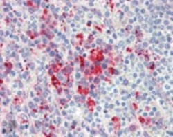
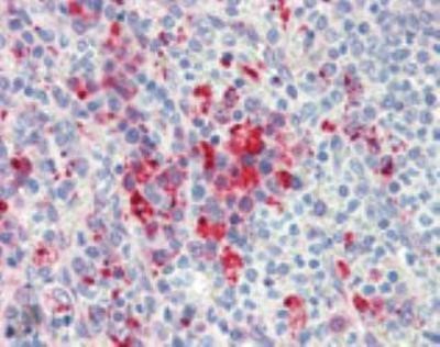
- Immunohistochemistry: HSP90 Antibody (AC88) [NB100-1972] - Analysis of human spleen tissue stained with HSP90, mAb (AC88) at 10 ug/ml.
Supportive validation
- Submitted by
- Novus Biologicals (provider)
- Main image

- Experimental details
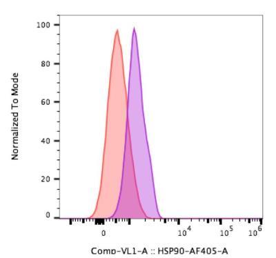
- Flow Cytometry: HSP90 Antibody (AC88) [NB100-1972] - Analysis using the Alexa Fluor 405 conjugated HSP90 antibody, NB100-1972AF405 (purple) and fluorescence minus one control (orange). Staining of HSP90 in Primary CD4+ T-cells derived from human peripheral blood using 1uL of antibody per 1X10e5 cells. Image from verified customer review.
- Submitted by
- Novus Biologicals (provider)
- Main image

- Experimental details
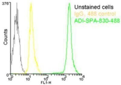
- Flow Cytometry: HSP90 Antibody (AC88) [NB100-1972] - Analysis using the DyLight 488 conjugate of NB100-1972. Staining of 106 Jurkat cells using HSP90 mAb (AC88), DyLight 488 Conjugate at a concentration of 50ug/ml.
- Submitted by
- Novus Biologicals (provider)
- Main image

- Experimental details
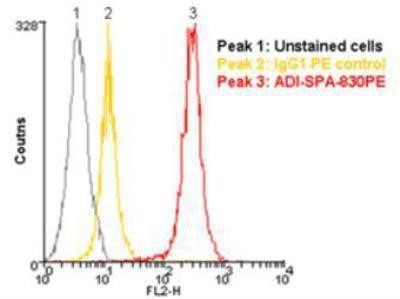
- Flow Cytometry: HSP90 Antibody (AC88) [NB100-1972] - Analysis using the PE conjugate of NB100-1972. Staining of 10^6 Jurkat cells using HSP90 mAb (AC88), R-Phycoerythrin Conjugate at a concentration of 10ug/mL
- Submitted by
- Novus Biologicals (provider)
- Main image
- Experimental details
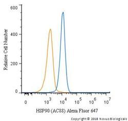
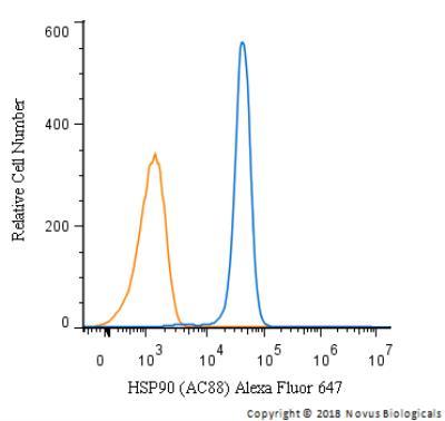
- Flow Cytometry: HSP90 Antibody (AC88) [NB100-1972] - An intracellular stain was performed on HeLa cells with HSP90 Antibody (AC88) NB100-1972AF647 (blue) and a matched isotype control (orange). Cells were fixed with 4% PFA and then permeabilized with 0.1% saponin. Cells were incubated in an antibody dilution of 2.5 ug/mL for 30 minutes at room temperature. Both antibodies were conjugated to Alexa Fluor 647.
- Submitted by
- Novus Biologicals (provider)
- Main image

- Experimental details
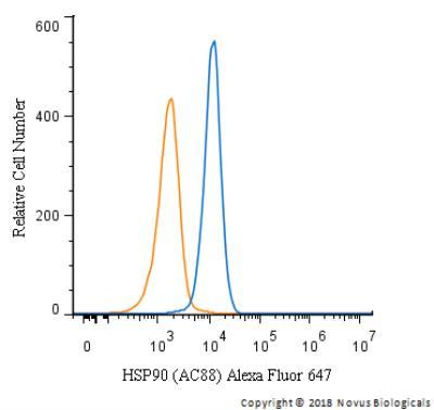
- Flow Cytometry: HSP90 Antibody (AC88) [NB100-1972] - An intracellular stain was performed on Jurkat cells with HSP90 Antibody (AC88) NB100-1972AF647 (blue) and a matched isotype control (orange). Cells were fixed with 4% PFA and then permeabilized with 0.1% saponin. Cells were incubated in an antibody dilution of 5 ug/mL for 30 minutes at room temperature. Both antibodies were conjugated to Alexa Fluor 647.
- Submitted by
- Novus Biologicals (provider)
- Main image
- Experimental details
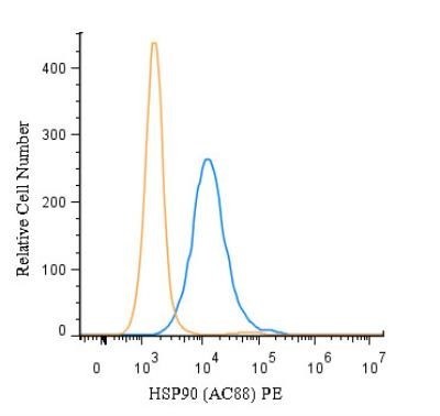
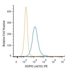
- Flow (Intracellular): HSP90 Antibody (AC88) [NB100-1972] - An intracellular stain was performed on HeLa cells with HSP90 Antibody (AC88) NB100-1972PE (blue) and a matched isotype control (orange). Cells were fixed with 4% PFA and then permeablized with 0.1% saponin. Cells were incubated in an antibody dilution of 5 ug/mL for 30 minutes at room temperature. Both antibodies were conjugated to phycoerythrin. Image using the PE format of this antibody.