Explore
 Validate
 Learn
Learn Western blot
 Immunocytochemistry
Antibody data
- Antibody Data
- Antigen structure
- References [2]
- Comments [0]
- Validations
- Western blot [2]
- Other assay [1]
Submit
Validation data
Reference
Comment
Report error
- Product number
- NBP2-50028 - Provider product page

- Provider
- Novus Biologicals
- Product name
- Chicken Polyclonal Calbindin D-28K Antibody
- Antibody type
- Polyclonal
- Description
- Ammonium sulfate precipitation.
- Reactivity
- Human, Mouse, Rat, Bovine
- Host
- Chicken/Avian
- Isotype
- IgY
- Vial size
- 0.1 ml
- Storage
- Store at 4C short term. Aliquot and store at -20C long term. Avoid freeze-thaw cycles.
Submitted references LKB1 coordinates neurite remodeling to drive synapse layer emergence in the outer retina.
Nonmuscle myosin 2 proteins encoded by Myh9, Myh10, and Myh14 are uniquely distributed in the tubular segments of murine kidney.
Burger CA, Alevy J, Casasent AK, Jiang D, Albrecht NE, Liang JH, Hirano AA, Brecha NC, Samuel MA
eLife 2020 May 7;9
eLife 2020 May 7;9
Nonmuscle myosin 2 proteins encoded by Myh9, Myh10, and Myh14 are uniquely distributed in the tubular segments of murine kidney.
Otterpohl KL, Hart RG, Evans C, Surendran K, Chandrasekar I
Physiological reports 2017 Dec;5(23)
Physiological reports 2017 Dec;5(23)
No comments: Submit comment
Supportive validation
- Submitted by
- Novus Biologicals (provider)
- Main image

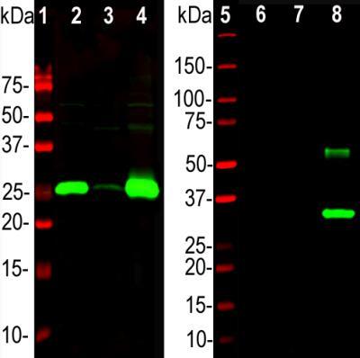
- Experimental details
- Western Blot: Calbindin D-28K Antibody [NBP2-50028] - Analysis of rat brain lysate (left), 0.5 ug of His-tagged recombinant proteins (right) were probed with NBP2-50028 at 1:5,000. Lane 1: Parvalbumin, Lane 2: Calretinin, Lane 3: Calbindin.

- Submitted by
- Novus Biologicals (provider)
- Main image

- Experimental details
- Western Blot: Calbindin D-28K Antibody [NBP2-50028] - Analysis of different tissue lysates and recombinant protein solutions using chicken pAb to calbindin, NBP2-50028, dilution 1:5,000 in green: [1] protein standard (red), [2] rat cerebellum, [3] pig hippocampus, [4] cow cerebellum, [5] protein standard (red). Next lanes are full length recombinant human proteins, [6] Parvalbumin, [7] Calretinin, [8] Calbindin. Bands at 25kDa in tissue lysates and ~30 kDa in protein solutions correspond to calbindin, the recombinant form being slightly larger due to the presence of a His tag and other vector derived sequence. NBP2-50028 antibody specifically recognizes calbindin protein and does not react with the closely related proteins parvalbumin and calretinin.
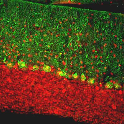
Supportive validation
- Submitted by
- Novus Biologicals (provider)
- Main image
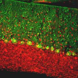
- Experimental details
- Immunohistochemistry Free-Floating: Calbindin D-28K Antibody [NBP2-50028] - Analysis of rat cerebellum section stained with chicken Calbindin D-28K pAb, dilution 1:2,000 (Green), and costained with rabbit MeCP2 pAb, dilution 1:5,000 (Red). Following transcardial perfusion with 4% paraformaldehyde, brain was post fixed for 24 hours, cut to 45uM, and free-floating sections were stained with above antibodies. Calbindin D-28K, often used as a Purkinje cell marker, is prominently detected in dendrites and perikarya of these cells in the cerebellar molecular layer. The MeCP2 antibody selectively stains nuclei of neuronal cells.