 Explore
 Validate
 Learn
Learn Western blot
 Immunocytochemistry
Antibody data
- Antibody Data
- Antigen structure
- References [0]
- Comments [0]
- Validations
- Western blot [2]
- Other assay [1]
Submit
Validation data
Reference
Comment
Report error
- Product number
- NBP2-50050 - Provider product page

- Provider
- Novus Biologicals
- Product name
- Mouse Monoclonal Calbindin D-28K Antibody
- Antibody type
- Monoclonal
- Description
- Immunogen affinity purified.
- Reactivity
- Human, Mouse, Rat, Bovine
- Host
- Mouse
- Isotype
- IgG
- Vial size
- 0.1 ml
- Concentration
- 1 mg/ml
- Storage
- Store at 4C short term. Aliquot and store at -20C long term. Avoid freeze-thaw cycles.
No comments: Submit comment
Supportive validation
- Submitted by
- Novus Biologicals (provider)
- Main image
- Experimental details
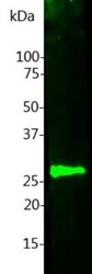
- Western Blot: Calbindin D-28K Antibody (5A9) [NBP2-50050] - Analysis of cow cerebellum lysate were probed with NBP2-50050. NBP2-50050 binds strongly and cleanly to Calbindin at 28 kDa.

- Submitted by
- Novus Biologicals (provider)
- Main image
- Experimental details
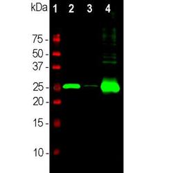
- Western Blot: Calbindin D-28K Antibody (5A9) [NBP2-50050] - Analysis of different neuronal tissue lysates using mouse mAb to Calbindin D-28K, dilution 1:5,000: [1] protein standard, [2] rat cerebellum, [3] pig hippocampus, and [4] cow cerebellum. Bands at about 26kDa corresponds to the calbindin protein, heavily expressed in the cerebellum but a very minor component of hippocampus.

Supportive validation
- Submitted by
- Novus Biologicals (provider)
- Main image

- Experimental details
- Immunohistochemistry Free-Floating: Calbindin D-28K Antibody (5A9) [NBP2-50050] - Analysis of rat brain cerebellum section stained with mouse mAb to Calbindin D-28K, dilution 1:2,000 (Red), and costained with rabbit GFAP pAb, dilution 1:5,000 (Green). DAPI staining of nuclear DNA (Blue). Following transcardial perfusion with 4% paraformaldehyde, brain was post fixed for 24hrs, cut to 45uM, and free-floating sections were stained with the above antibodies. The Calbindin D-28K antibody prominently labels the dendrites and perikarya of Purkinje cells in the molecular layer of the cerebellum. The GFAP antibody stains the processes of Bergmann glia in the molecular layer and astroglia in the granular and white matter layers of cerebellum.
