 Explore
 Validate
 Learn
Learn Western blot
Antibody data
- Antibody Data
- Antigen structure
- References [0]
- Comments [0]
- Validations
- Western blot [1]
- Immunohistochemistry [1]
- Flow cytometry [1]
Submit
Validation data
Reference
Comment
Report error
- Product number
- MAB6480 - Provider product page

- Provider
- Novus Biologicals
- Product name
- Rat Monoclonal CEACAM1/CD66a Antibody
- Antibody type
- Monoclonal
- Description
- Protein A or G purified from hybridoma culture supernatant. Detects mouse CEACAM-1/CD66a in direct ELISAs and Western blots. In direct ELISAs, no cross-reactivity with recombinant human CECAM-1, -3, -5, or -6 is observed. In Western blots, no cross-reactivity with rhCEACAM-1, -3, -4, -5, -6, or -7 is observed.
- Reactivity
- Mouse
- Host
- Rat
- Conjugate
- Unconjugated
- Isotype
- IgG
- Vial size
- 100 ug
- Concentration
- LYOPH
- Storage
- Use a manual defrost freezer and avoid repeated freeze-thaw cycles. 12 months from date of receipt, -20 to -70 degreesC as supplied. 1 month, 2 to 8 degreesC under sterile conditions after reconstitution. 6 months, -20 to -70 degreesC under sterile conditions after reconstitution.
No comments: Submit comment
Supportive validation
- Submitted by
- Novus Biologicals (provider)
- Main image
- Experimental details
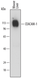
- Detection of Mouse CEACAM-1/CD66a by Western Blot. Western blot shows lysates of mouse liver tissue. PVDF membrane was probed with 0.1 µg/mL of Rat Anti-Mouse CEACAM-1/CD66a Monoclonal Antibody (Catalog # MAB6480) followed by HRP-conjugated Anti-Rat IgG Secondary Antibody (Catalog # HAF005). A specific band was detected for CEACAM-1/CD66a at approximately 110-120 kDa (as indicated). This experiment was conducted under reducing conditions and using Immunoblot Buffer Group 1.

Supportive validation
- Submitted by
- Novus Biologicals (provider)
- Main image

- Experimental details
- CEACAM-1/CD66a in Mouse Liver. CEACAM-1/CD66a was detected in perfusion fixed frozen sections of mouse liver using Rat Anti-Mouse CEACAM-1/CD66a Monoclonal Antibody (Catalog # MAB6480) at 25 µg/mL overnight at 4 °C. Tissue was stained using the NorthernLights™ 557-conjugated Anti-Rat IgG Secondary Antibody (red; Catalog # NL013) and counterstained with DAPI (blue). Specific staining was localized to bile canaliculi. View our protocol for Fluorescent IHC Staining of Frozen Tissue Sections.

Supportive validation
- Submitted by
- Novus Biologicals (provider)
- Main image

- Experimental details
- Detection of CEACAM-1 in Mouse Splenocytes by Flow Cytometry. Mouse splenocytes were stained with Rat Anti-Mouse CEACAM-1/CD66a Monoclonal Antibody (Catalog # MAB6480) followed by Fluorescein-conjugated Anti-Rat IgG Secondary Antibody (Catalog # F0104B) and Rat Anti-Mouse B220/CD45R Allophycocyanin-conjugated Monoclonal Antibody (Catalog # FAB1217A). Quadrant markers were set based on control antibody staining (Catalog # MAB005).
