 Explore
 Validate
 Learn
Learn Western blot
 Immunoprecipitation
Antibody data
- Antibody Data
- Antigen structure
- References [0]
- Comments [0]
- Validations
- Western blot [1]
- ELISA [1]
- Immunocytochemistry [1]
- Immunohistochemistry [1]
- Flow cytometry [1]
Submit
Validation data
Reference
Comment
Report error
- Product number
- AM26012PU-N - Provider product page

- Provider
- Acris Antibodies GmbH
- Proper citation
- Acris Antibodies GmbH Cat#AM26012PU-N, RRID:AB_10849032
- Product name
- anti CD66a / CEACAM1
- Antibody type
- Monoclonal
- Antigen
- Recombinant Human soluble CEACAM-1-Fc lacking the N-domain (produced in HEK293 cells)
- Reactivity
- Human
- Host
- Mouse
- Isotype
- IgG
- Antibody clone number
- B3
- Vial size
- 0.1 mg
No comments: Submit comment
Supportive validation
- Submitted by
- Acris Antibodies GmbH (provider)
- Main image
- Experimental details
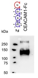
- Western blot analysis: Human CEACAM-1 lysate Detection utilysing 10 μg/ml

Supportive validation
- Submitted by
- Acris Antibodies GmbH (provider)
- Main image
- Experimental details
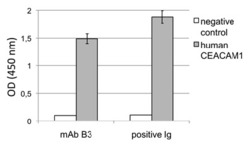
- Sandwich ELISA: Solid phase was coated with 3 μg/ml anti CEA binding human CEACAM1- CEACAM8. After washing, blocking and coating human CEACAM1 antigen, detecting antibody mAb B3 (10 μg/ml) followed by HRP-coupled goat anti-mouse Ig was added. TMB was used for visualizing the binding measured by Tecan-ELISA reader at 450 nm.

Supportive validation
- Submitted by
- Acris Antibodies GmbH (provider)
- Main image

- Experimental details
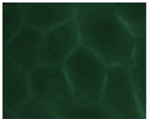
- Immunofluorescence with HeLa-CEACAM-1 cells. Cells were 4% PFA fixed (10min) and then incubated in 5% BSA/PBS for 1 h to block non-specific prtein-protein interactions. The cells were then incubated with 10μg B3-17 overnight at 4°C. The secondary antibody (green) was Alexa Flour®488 goat-anti-Mouse IgG (H+L).
Supportive validation
- Submitted by
- Acris Antibodies GmbH (provider)
- Main image
- Experimental details
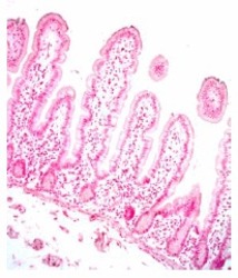
- IHC staining of human jejunum tissue with Monoclonal Antibody B3. CEACAM1 was detected in PFA-fixed paraffin-embedded sections of human jejunum tissue using B3 followed by staining with anti-mouse HRP-DAB and counterstaining with hematoxylin. The labeling showed week staining of CEACAM1 by B3.

Supportive validation
- Submitted by
- Acris Antibodies GmbH (provider)
- Main image
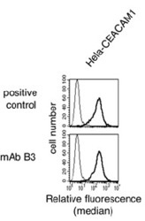
- Experimental details
- Flow cytometry: 10 μg/ml of primary mAb B3; 250.000 Hela-human CEACAM1 cells
