 Explore
 Validate
 Learn
Learn Western blot
Antibody data
- Antibody Data
- Antigen structure
- References [0]
- Comments [0]
- Validations
- Western blot [1]
- Immunohistochemistry [3]
Submit
Validation data
Reference
Comment
Report error
- Product number
- SP5403 - Provider product page

- Provider
- Acris Antibodies GmbH
- Proper citation
- Acris Antibodies GmbH Cat#SP5403, RRID:AB_1001072
- Product name
- anti NOS1
- Antibody type
- Polyclonal
- Antigen
- Synthetic Peptide corresponding to amino acid residues 724-739 of Rat bNOS.
- Reactivity
- Mouse, Rat, Bovine, Rabbit
- Host
- Rabbit
- Vial size
- 0.1 ml
No comments: Submit comment
Supportive validation
- Submitted by
- Acris Antibodies GmbH (provider)
- Main image

- Experimental details
- Western blot analysis of nNOS was performed by loading 40 µg of Mouse (Lane 1) and Rat Brain (Lane 2) tissue lysate onto a 4-12% Bis-Tris polyacrylamide gel. Proteins were transferred to a Nitrocellulose membrane. Membranes were probed with a Rabbit polyclonal antibody Cat.-No SP5403 recognizing nNOS at a dilution of 1/1000. The western blot was performed using the Fast Western Kit supersignal West Dura.

Supportive validation
- Submitted by
- Acris Antibodies GmbH (provider)
- Main image

- Experimental details
- Immunohistochemistry was performed on normal biopsies of deparaffinized Rat cerebellum tissue. To expose target proteins, heat induced antigen retrieval was performed using 10mM sodium citrate (pH6.0) buffer, microwaved for 8-15 minutes. Following antigen retrieval tissues were blocked in 3% BSA-PBS for 30 minutes at room temperature. Tissues were then probed at a dilution of 1/200 with Cat.-No SP5403 or without primary antibody (negative control) overnight at 4°C in a humidified chamber.

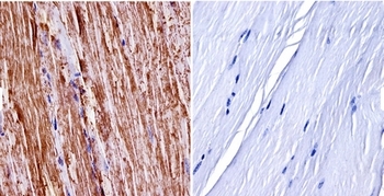
- Submitted by
- Acris Antibodies GmbH (provider)
- Main image

- Experimental details
- Immunohistochemistry was performed on normal biopsies of deparaffinized mouse skeletal muscle tissue. To expose target proteins, heat induced antigen retrieval was performed using 10mM sodium citrate (pH6.0) buffer, microwaved for 8-15 minutes. Following antigen retrieval tissues were blocked in 3% BSA-PBS for 30 minutes at room temperature. Tissues were then probed at a dilution of 1/400 with Cat.-No SP5403 or without primary antibody (negative control) overnight at 4°C in a humidified chamber.
- Submitted by
- Acris Antibodies GmbH (provider)
- Main image
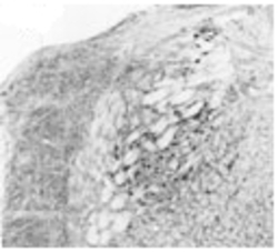
- Experimental details
- Immunohistochemical staining of bNOS in rat brain using SP5403 antibody.
