Explore
 Validate
 Learn
Learn ELISA
ELISAAntibody data
- Antibody Data
- Antigen structure
- References [16]
- Comments [0]
- Validations
- ELISA [1]
- Immunocytochemistry [1]
- Immunohistochemistry [2]
- Blocking/Neutralizing [1]
Submit
Validation data
Reference
Comment
Report error
- Product number
- AF1433 - Provider product page

- Provider
- R&D Systems
- Product name
- Human Osteopontin/OPN Antibody
- Antibody type
- Polyclonal
- Description
- Antigen Affinity-purified. Detects human Osteopontin/OPN in direct ELISAs and Western blots. In direct ELISAs, less than 10% cross-reactivity with recombinant rat Osteopontin and recombinant mouse OPN is observed.
- Reactivity
- Human
- Host
- Goat
- Conjugate
- Unconjugated
- Isotype
- IgG
- Vial size
- 100 ug
- Concentration
- LYOPH
- Storage
- Use a manual defrost freezer and avoid repeated freeze-thaw cycles. 12 months from date of receipt, -20 to -70 °C as supplied. 1 month, 2 to 8 °C under sterile conditions after reconstitution. 6 months, -20 to -70 °C under sterile conditions after reconstitution.
Submitted references Osteopontin mediates glioblastoma-associated macrophage infiltration and is a potential therapeutic target.
Differential expression patterns of Toll Like Receptors and Interleukin-37 between calcific aortic and mitral valve cusps in humans.
Single-Cell RNA-Seq Reveals the Transcriptional Landscape and Heterogeneity of Aortic Macrophages in Murine Atherosclerosis.
Phenotypic and Functional Characterization of Peripheral Sensory Neurons derived from Human Embryonic Stem Cells.
Osteopontin contributes to effective neutrophil recruitment, IL-1β production and apoptosis in Aspergillus fumigatus keratitis.
Generation of iPSC-derived limb progenitor-like cells for stimulating phalange regeneration in the adult mouse.
Osteopontin-metallothionein I/II interactions in experimental autoimmune encephalomyelitis.
Inhibition of Cellular Adhesion by Immunological Targeting of Osteopontin Neoepitopes Generated through Matrix Metalloproteinase and Thrombin Cleavage.
Cell stress induces upregulation of osteopontin via the ERK pathway in type II alveolar epithelial cells.
Osteopontin promotes the invasive growth of melanoma cells by activating integrin αvβ3 and down-regulating tetraspanin CD9.
CD44v6 is a marker of constitutive and reprogrammed cancer stem cells driving colon cancer metastasis.
Mechanism of hepatitis C virus (HCV)-induced osteopontin and its role in epithelial to mesenchymal transition of hepatocytes.
Regulation and function of immunosuppressive molecule human leukocyte antigen G5 in human bone tissue.
Osteopontin is a prognostic factor for survival of acute myeloid leukemia patients.
Secreted osteopontin is highly polymerized in human airways and fragmented in asthmatic airway secretions.
The crucial role of cyclooxygenase-2 in osteopontin-induced protein kinase C alpha/c-Src/IkappaB kinase alpha/beta-dependent prostate tumor progression and angiogenesis.
Wei J, Marisetty A, Schrand B, Gabrusiewicz K, Hashimoto Y, Ott M, Grami Z, Kong LY, Ling X, Caruso H, Zhou S, Wang YA, Fuller GN, Huse J, Gilboa E, Kang N, Huang X, Verhaak R, Li S, Heimberger AB
The Journal of clinical investigation 2019 Jan 2;129(1):137-149
The Journal of clinical investigation 2019 Jan 2;129(1):137-149
Differential expression patterns of Toll Like Receptors and Interleukin-37 between calcific aortic and mitral valve cusps in humans.
Kapelouzou A, Kontogiannis C, Tsilimigras DI, Georgiopoulos G, Kaklamanis L, Tsourelis L, Cokkinos DV
Cytokine 2019 Apr;116:150-160
Cytokine 2019 Apr;116:150-160
Single-Cell RNA-Seq Reveals the Transcriptional Landscape and Heterogeneity of Aortic Macrophages in Murine Atherosclerosis.
Cochain C, Vafadarnejad E, Arampatzi P, Pelisek J, Winkels H, Ley K, Wolf D, Saliba AE, Zernecke A
Circulation research 2018 Jun 8;122(12):1661-1674
Circulation research 2018 Jun 8;122(12):1661-1674
Phenotypic and Functional Characterization of Peripheral Sensory Neurons derived from Human Embryonic Stem Cells.
Alshawaf AJ, Viventi S, Qiu W, D'Abaco G, Nayagam B, Erlichster M, Chana G, Everall I, Ivanusic J, Skafidas E, Dottori M
Scientific reports 2018 Jan 12;8(1):603
Scientific reports 2018 Jan 12;8(1):603
Osteopontin contributes to effective neutrophil recruitment, IL-1β production and apoptosis in Aspergillus fumigatus keratitis.
Zhao G, Hu M, Li C, Lee J, Yuan K, Zhu G, Che C
Immunology and cell biology 2018 Apr;96(4):401-412
Immunology and cell biology 2018 Apr;96(4):401-412
Generation of iPSC-derived limb progenitor-like cells for stimulating phalange regeneration in the adult mouse.
Chen Y, Xu H, Lin G
Cell discovery 2017;3:17046
Cell discovery 2017;3:17046
Osteopontin-metallothionein I/II interactions in experimental autoimmune encephalomyelitis.
Jakovac H, Grubić Kezele T, Šućurović S, Mulac-Jeričević B, Radošević-Stašić B
Neuroscience 2017 May 14;350:133-145
Neuroscience 2017 May 14;350:133-145
Inhibition of Cellular Adhesion by Immunological Targeting of Osteopontin Neoepitopes Generated through Matrix Metalloproteinase and Thrombin Cleavage.
Jürets A, Le Bras M, Staffler G, Stein G, Leitner L, Neuhofer A, Tardelli M, Turkof E, Zeyda M, Stulnig TM
PloS one 2016;11(2):e0148333
PloS one 2016;11(2):e0148333
Cell stress induces upregulation of osteopontin via the ERK pathway in type II alveolar epithelial cells.
Kato A, Okura T, Hamada C, Miyoshi S, Katayama H, Higaki J, Ito R
PloS one 2014;9(6):e100106
PloS one 2014;9(6):e100106
Osteopontin promotes the invasive growth of melanoma cells by activating integrin αvβ3 and down-regulating tetraspanin CD9.
Yin M, Soikkeli J, Jahkola T, Virolainen S, Saksela O, Hölttä E
The American journal of pathology 2014 Mar;184(3):842-58
The American journal of pathology 2014 Mar;184(3):842-58
CD44v6 is a marker of constitutive and reprogrammed cancer stem cells driving colon cancer metastasis.
Todaro M, Gaggianesi M, Catalano V, Benfante A, Iovino F, Biffoni M, Apuzzo T, Sperduti I, Volpe S, Cocorullo G, Gulotta G, Dieli F, De Maria R, Stassi G
Cell stem cell 2014 Mar 6;14(3):342-56
Cell stem cell 2014 Mar 6;14(3):342-56
Mechanism of hepatitis C virus (HCV)-induced osteopontin and its role in epithelial to mesenchymal transition of hepatocytes.
Iqbal J, McRae S, Banaudha K, Mai T, Waris G
The Journal of biological chemistry 2013 Dec 27;288(52):36994-7009
The Journal of biological chemistry 2013 Dec 27;288(52):36994-7009
Regulation and function of immunosuppressive molecule human leukocyte antigen G5 in human bone tissue.
Deschaseaux F, Gaillard J, Langonné A, Chauveau C, Naji A, Bouacida A, Rosset P, Heymann D, De Pinieux G, Rouas-Freiss N, Sensébé L
FASEB journal : official publication of the Federation of American Societies for Experimental Biology 2013 Aug;27(8):2977-87
FASEB journal : official publication of the Federation of American Societies for Experimental Biology 2013 Aug;27(8):2977-87
Osteopontin is a prognostic factor for survival of acute myeloid leukemia patients.
Liersch R, Gerss J, Schliemann C, Bayer M, Schwöppe C, Biermann C, Appelmann I, Kessler T, Löwenberg B, Büchner T, Hiddemann W, Müller-Tidow C, Berdel WE, Mesters R
Blood 2012 May 31;119(22):5215-20
Blood 2012 May 31;119(22):5215-20
Secreted osteopontin is highly polymerized in human airways and fragmented in asthmatic airway secretions.
Arjomandi M, Frelinger J, Donde A, Wong H, Yellamilli A, Raymond W
PloS one 2011;6(10):e25678
PloS one 2011;6(10):e25678
The crucial role of cyclooxygenase-2 in osteopontin-induced protein kinase C alpha/c-Src/IkappaB kinase alpha/beta-dependent prostate tumor progression and angiogenesis.
Jain S, Chakraborty G, Kundu GC
Cancer research 2006 Jul 1;66(13):6638-48
Cancer research 2006 Jul 1;66(13):6638-48
No comments: Submit comment
Supportive validation
- Submitted by
- R&D Systems (provider)
- Main image
- Experimental details
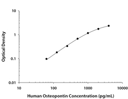
- Human Osteopontin/OPN ELISA Standard Curve. Recombinant Human Osteopontin/OPN protein was serially diluted 2-fold and captured by Mouse Anti-Human Osteopontin/OPN Monoclonal Antibody (Catalog # MAB14332R) coated on a Clear Polystyrene Microplate (Catalog # DY990). Goat Anti-Human Osteopontin/OPN Antigen Affinity-purified Polyclonal Antibody (Catalog # AF1433) was biotinylated and incubated with the protein captured on the plate. Detection of the standard curve was achieved by incubating Streptavidin-HRP (Catalog # DY998) followed by Substrate Solution (Catalog # DY999) and stopping the enzymatic reaction with Stop Solution (Catalog # DY994).

Supportive validation
- Submitted by
- R&D Systems (provider)
- Main image

- Experimental details
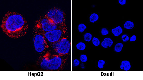
- Osteopontin/OPN in HepG2 Human Cell Line. Osteopontin/OPN was detected in immersion fixed HepG2 human hepatocellular carcinoma cell line (positive staining; left panel) and Daudi human Burkitt's lymphoma cell line (negative staining; right panel) using Goat Anti-Human Osteopontin/OPN Antigen Affinity-purified Polyclonal Antibody (Catalog # AF1433) at 1.7 µg/mL for 3 hours at room temperature. Cells were stained using the NorthernLights™ 557-conjugated Anti-Goat IgG Secondary Antibody (red; Catalog # NL001) and counterstained with DAPI (blue). Specific staining was localized to cytoplasm. View our protocol for Fluorescent ICC Staining of Cells on Coverslips.
Supportive validation
- Submitted by
- R&D Systems (provider)
- Main image
- Experimental details
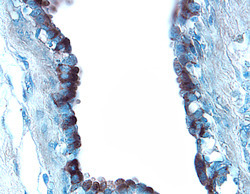
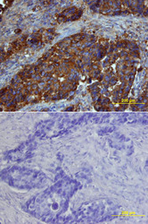
- Osteopontin/OPN in Human Breast Cancer Tissue. Osteopontin/OPN was detected in immersion fixed paraffin-embedded sections of human breast cancer tissue using 8 µg/mL Goat Anti-Human Osteopontin/OPN Antigen Affinity-purified Polyclonal Antibody (Catalog # AF1433) overnight at 4 °C. Tissue was stained with the Anti-Goat HRP-DAB Cell & Tissue Staining Kit (brown; Catalog # CTS008) and counterstained with hematoxylin (blue). Specific labeling was localized to the surface of epithelial cells in the intralobular duct. View our protocol for Chromogenic IHC Staining of Paraffin-embedded Tissue Sections.

- Submitted by
- R&D Systems (provider)
- Main image
- Experimental details
- Osteopontin/OPN in Human Breast Cancer Tissue. Osteopontin/OPN was detected in immersion fixed paraffin-embedded sections of human breast cancer tissue using Goat Anti-Human Osteopontin/OPN Antigen Affinity-purified Polyclonal Antibody (Catalog # AF1433) at 15 µg/mL overnight at 4 °C. Tissue was stained using the Anti-Goat HRP-DAB Cell & Tissue Staining Kit (brown; Catalog # CTS008) and counterstained with hematoxylin (blue). Lower panel shows a lack of labeling if primary antibodies are omitted and tissue is stained only with secondary antibody followed by incubation with detection reagents. View our protocol for Chromogenic IHC Staining of Paraffin-embedded Tissue Sections.

Supportive validation
- Submitted by
- R&D Systems (provider)
- Main image
- Experimental details
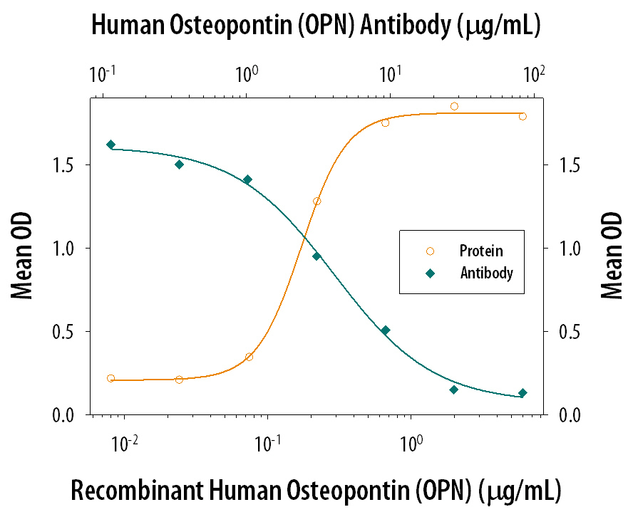
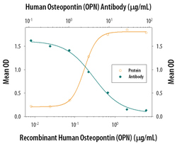
- Cell Adhesion Mediated by Osteopontin/OPN and Neutralization by Human Osteopontin/OPN Antibody. Recombinant Human Osteopontin/OPN (Catalog # 1433-OP), immobilized onto a microplate, supports the adhesion of the HEK293 human embryonic kidney cell line in a dose-dependent manner (orange line). Adhesion elicited by Recombinant Human Osteopontin/OPN (1 µg/mL) is neutralized (green line) by increasing concentrations of Goat Anti-Human Osteopontin/OPN Antigen Affinity-purified Polyclonal Antibody (Catalog # AF1433). The ND50 is typically 2-6 µg/mL.