Explore
 Validate
 Learn
Learn Western blot
 Immunocytochemistry
Antibody data
- Antibody Data
- Antigen structure
- References [1]
- Comments [0]
- Validations
- Western blot [3]
- Immunohistochemistry [1]
Submit
Validation data
Reference
Comment
Report error
- Product number
- AF1565 - Provider product page

- Provider
- R&D Systems
- Product name
- Human/Mouse/Rat FABP1/L-FABP Antibody
- Antibody type
- Polyclonal
- Description
- Antigen Affinity-purified. Detects rat FABP1/L-FABP in direct ELISAs and Western blots.
- Reactivity
- Human, Mouse, Rat
- Host
- Goat
- Conjugate
- Unconjugated
- Antigen sequence
P02692- Isotype
- IgG
- Vial size
- 100 ug
- Concentration
- LYOPH
- Storage
- Use a manual defrost freezer and avoid repeated freeze-thaw cycles. 12 months from date of receipt, -20 to -70 °C as supplied. 1 month, 2 to 8 °C under sterile conditions after reconstitution. 6 months, -20 to -70 °C under sterile conditions after reconstitution.
Submitted references Identification of biomarkers associated with the development of hepatocellular carcinoma in CuZn superoxide dismutase deficient mice.
Elchuri S, Naeemuddin M, Sharpe O, Robinson WH, Huang TT
Proteomics 2007 Jun;7(12):2121-9
Proteomics 2007 Jun;7(12):2121-9
No comments: Submit comment
Supportive validation
- Submitted by
- R&D Systems (provider)
- Main image
- Experimental details
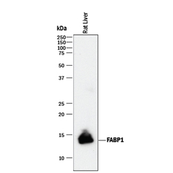
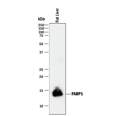
- Detection of Rat FABP1/L-FABP by Western Blot. Western blot shows lysate of rat liver tissue. PVDF membrane was probed with 0.2 µg/mL of Goat Anti-Human/Mouse/ Rat FABP1/L-FABP Antigen Affinity-purified Polyclonal Antibody (Catalog # AF1565) followed by HRP-conjugated Anti-Goat IgG Secondary Antibody (Catalog # HAF017). A specific band was detected for FABP1/ L-FABP at approximately 13 kDa (as indicated). This experiment was conducted under reducing conditions and using Immunoblot Buffer Group 1.
- Submitted by
- R&D Systems (provider)
- Main image
- Experimental details
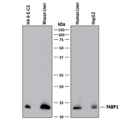
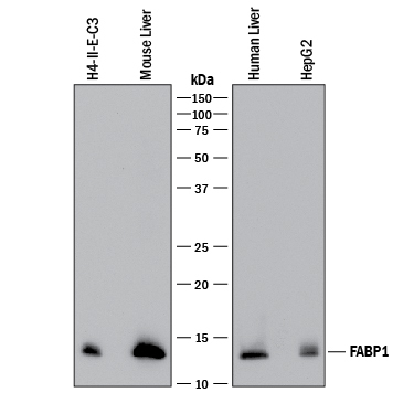
- Detection of Human, Mouse, and Rat FABP1/L-FABP by Western Blot. Western blot shows lysates of H4-II-E-C3 rat hepatoma cell line, mouse liver tissue, human liver tissue, and HepG2 human hepatocellular carcinoma cell line. PVDF membrane was probed with 0.5 µg/mL of Goat Anti-Human/ Mouse/Rat FABP1/L-FABP Antigen Affinity-purified Polyclonal Antibody (Catalog # AF1565) followed by HRP-conjugated Anti-Goat IgG Secondary Antibody (Catalog # HAF017). A specific band was detected for FABP1/L-FABP at approximately 13 kDa (as indicated). This experiment was conducted under reducing conditions and using Immunoblot Buffer Group 1.
- Submitted by
- R&D Systems (provider)
- Main image

- Experimental details
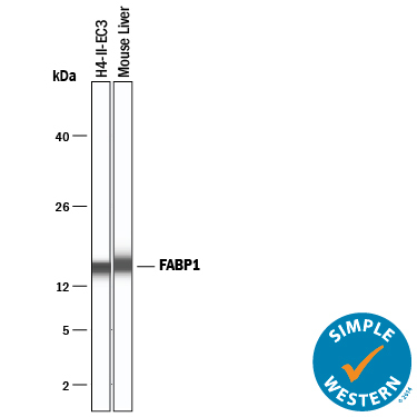
- Detection of Mouse and Rat FABP1/L-FABP by Simple WesternTM. Simple Western lane view shows lysates of H4-II-E-C3 rat hepatoma cell line and mouse liver tissue, loaded at 0.2 mg/mL. A specific band was detected for FABP1/L-FABP at approximately 16 kDa (as indicated) using 10 µg/mL of Goat Anti-Human/Mouse/Rat FABP1/L-FABP Antigen Affinity-purified Polyclonal Antibody (Catalog # AF1565) followed by 1:50 dilution of HRP-conjugated Anti-Goat IgG Secondary Antibody (Catalog # HAF109). This experiment was conducted under reducing conditions and using the 12-230 kDa separation system.
Supportive validation
- Submitted by
- R&D Systems (provider)
- Main image

- Experimental details
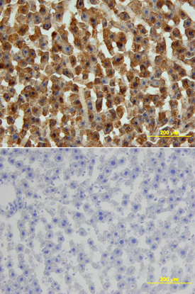
- FABP1 in Rat Liver. FABP1 was detected in perfusion fixed frozen sections of rat liver using Goat Anti-Human/Mouse/Rat FABP1 Antigen Affinity-purified Polyclonal Antibody (Catalog # AF1565) at 15 µg/mL overnight at 4 °C. Tissue was stained using the Anti-Goat HRP-DAB Cell & Tissue Staining Kit (brown; Catalog # CTS008) and counterstained with hematoxylin (blue). Lower panel shows a lack of labeling if primary antibodies are omitted and tissue is stained only with secondary antibody followed by incubation with detection reagents. View our protocol for Chromogenic IHC Staining of Frozen Tissue Sections.