Antibody data
- Antibody Data
- Antigen structure
- References [4]
- Comments [0]
- Validations
- Western blot [1]
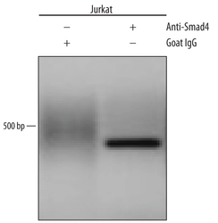
- Chromatin Immunoprecipitation [1]
Submit
Validation data
Reference
Comment
Report error
- Product number
- AF2097 - Provider product page

- Provider
- Novus Biologicals
- Product name
- Goat Polyclonal Smad4 Antibody
- Antibody type
- Polyclonal
- Description
- Immunogen affinity purified. Detects human Smad4 in direct ELISAs and Western blots. In direct ELISAs and Western blots, approximately 15% cross-reactivity with recombinant human (rh) Smad1 and less than 5% cross-reactivity with rhSmad9, rhSmad5, and rhSmad7 is observed.
- Reactivity
- Human
- Host
- Goat
- Conjugate
- Unconjugated
- Isotype
- IgG
- Vial size
- 100 ug
- Concentration
- LYOPH
- Storage
- Use a manual defrost freezer and avoid repeated freeze-thaw cycles. 12 months from date of receipt, -20 to -70 degreesC as supplied. 1 month, 2 to 8 degreesC under sterile conditions after reconstitution. 6 months, -20 to -70 degreesC under sterile conditions after reconstitution.
Submitted references Characterization of Membrane Integrity and Morphological Stability of Human Salivary Exosomes.
SMAD3 and SMAD4 have a more dominant role than SMAD2 in TGFβ-induced chondrogenic differentiation of bone marrow-derived mesenchymal stem cells.
An interaction network of mental disorder proteins in neural stem cells.
Chromatin and transcriptional signatures for Nodal signaling during endoderm formation in hESCs.
Kumeda N, Ogawa Y, Akimoto Y, Kawakami H, Tsujimoto M, Yanoshita R
Biological & pharmaceutical bulletin 2017;40(8):1183-1191
Biological & pharmaceutical bulletin 2017;40(8):1183-1191
SMAD3 and SMAD4 have a more dominant role than SMAD2 in TGFβ-induced chondrogenic differentiation of bone marrow-derived mesenchymal stem cells.
de Kroon LM, Narcisi R, van den Akker GG, Vitters EL, Blaney Davidson EN, van Osch GJ, van der Kraan PM
Scientific reports 2017 Feb 27;7:43164
Scientific reports 2017 Feb 27;7:43164
An interaction network of mental disorder proteins in neural stem cells.
Moen MJ, Adams HH, Brandsma JH, Dekkers DH, Akinci U, Karkampouna S, Quevedo M, Kockx CE, Ozgür Z, van IJcken WF, Demmers J, Poot RA
Translational psychiatry 2017 Apr 4;7(4):e1082
Translational psychiatry 2017 Apr 4;7(4):e1082
Chromatin and transcriptional signatures for Nodal signaling during endoderm formation in hESCs.
Kim SW, Yoon SJ, Chuong E, Oyolu C, Wills AE, Gupta R, Baker J
Developmental biology 2011 Sep 15;357(2):492-504
Developmental biology 2011 Sep 15;357(2):492-504
No comments: Submit comment
Supportive validation
- Submitted by
- Novus Biologicals (provider)
- Main image

- Experimental details
- Detection of Human Smad4 by Western Blot. Western blot shows lysates of HeLa human cervical epithelial carcinoma cell line, Jurkat human acute T cell leukemia cell line, and K562 human chronic myelogenous leukemia cell line. PVDF membrane was probed with 0.25 µg/mL of Goat Anti-Human Smad4 Antigen Affinity-purified Polyclonal Antibody (Catalog # AF2097) followed by HRP-conjugated Anti-Goat IgG Secondary Antibody (Catalog # HAF019). A specific band was detected for Smad4 at approximately 60 kDa (as indicated). This experiment was conducted under reducing conditions and using Immunoblot Buffer Group 1.
Supportive validation
- Submitted by
- Novus Biologicals (provider)
- Main image

- Experimental details
- Detection of Smad4-regulated Genes by Chromatin Immunoprecipitation. Jurkat human acute T cell leukemia cell line treated with 10 ng/mL Recombinant Human IL-12 (Catalog # 219-IL) overnight was fixed using formaldehyde, resuspended in lysis buffer, and sonicated to shear chromatin. Smad4/DNA complexes were immunoprecipitated using 5 μg Goat Anti-Human Smad4 Antigen Affinity-purified Polyclonal Antibody (Catalog # AF2097) or control antibody (Catalog # AB-108-C) for 15 minutes in an ultrasonic bath, followed by Biotinylated Anti-Goat IgG Secondary Antibody (Catalog # BAF109). Immunocomplexes were captured using 50 μL of MagCellect Streptavidin Ferrofluid (Catalog # MAG999) and DNA was purified using chelating resin solution. The p21 promoter was detected by standard PCR.
 Explore
 Validate
 Learn
Learn Western blot
 Immunocytochemistry