Explore
 Validate
 Learn
Learn Western blot
 Immunocytochemistry
Antibody data
- Antibody Data
- Antigen structure
- References [10]
- Comments [0]
- Validations
- Immunocytochemistry [1]
- Immunohistochemistry [1]
Submit
Validation data
Reference
Comment
Report error
- Product number
- AF2335 - Provider product page

- Provider
- R&D Systems
- Product name
- Mouse Aminopeptidase N/CD13 Antibody
- Antibody type
- Polyclonal
- Description
- Antigen Affinity-purified. Detects mouse Aminopeptidase N/CD13 in direct ELISAs and Western blots.
- Reactivity
- Mouse
- Host
- Goat
- Conjugate
- Unconjugated
- Antigen sequence
P97449- Isotype
- IgG
- Vial size
- 100 ug
- Concentration
- LYOPH
- Storage
- Use a manual defrost freezer and avoid repeated freeze-thaw cycles. 12 months from date of receipt, -20 to -70 °C as supplied. 1 month, 2 to 8 °C under sterile conditions after reconstitution. 6 months, -20 to -70 °C under sterile conditions after reconstitution.
Submitted references Single-cell transcriptomes of the regenerating intestine reveal a revival stem cell.
Microglia are an essential component of the neuroprotective scar that forms after spinal cord injury.
Endothelial Calcineurin Signaling Restrains Metastatic Outgrowth by Regulating Bmp2.
Required growth facilitators propel axon regeneration across complete spinal cord injury.
Apoε4 disrupts neurovascular regulation and undermines white matter integrity and cognitive function.
Blood-brain barrier-associated pericytes internalize and clear aggregated amyloid-β42 by LRP1-dependent apolipoprotein E isoform-specific mechanism.
Regional early and progressive loss of brain pericytes but not vascular smooth muscle cells in adult mice with disrupted platelet-derived growth factor receptor-β signaling.
Prostacyclin prevents pericyte loss and demyelination induced by lysophosphatidylcholine in the central nervous system.
Adiponectin expression protects against angiotensin II-mediated inflammation and accelerated atherosclerosis.
Pericyte loss influences Alzheimer-like neurodegeneration in mice.
Ayyaz A, Kumar S, Sangiorgi B, Ghoshal B, Gosio J, Ouladan S, Fink M, Barutcu S, Trcka D, Shen J, Chan K, Wrana JL, Gregorieff A
Nature 2019 May;569(7754):121-125
Nature 2019 May;569(7754):121-125
Microglia are an essential component of the neuroprotective scar that forms after spinal cord injury.
Bellver-Landete V, Bretheau F, Mailhot B, Vallières N, Lessard M, Janelle ME, Vernoux N, Tremblay MÈ, Fuehrmann T, Shoichet MS, Lacroix S
Nature communications 2019 Jan 31;10(1):518
Nature communications 2019 Jan 31;10(1):518
Endothelial Calcineurin Signaling Restrains Metastatic Outgrowth by Regulating Bmp2.
Hendrikx S, Coso S, Prat-Luri B, Wetterwald L, Sabine A, Franco CA, Nassiri S, Zangger N, Gerhardt H, Delorenzi M, Petrova TV
Cell reports 2019 Jan 29;26(5):1227-1241.e6
Cell reports 2019 Jan 29;26(5):1227-1241.e6
Required growth facilitators propel axon regeneration across complete spinal cord injury.
Anderson MA, O'Shea TM, Burda JE, Ao Y, Barlatey SL, Bernstein AM, Kim JH, James ND, Rogers A, Kato B, Wollenberg AL, Kawaguchi R, Coppola G, Wang C, Deming TJ, He Z, Courtine G, Sofroniew MV
Nature 2018 Sep;561(7723):396-400
Nature 2018 Sep;561(7723):396-400
Apoε4 disrupts neurovascular regulation and undermines white matter integrity and cognitive function.
Koizumi K, Hattori Y, Ahn SJ, Buendia I, Ciacciarelli A, Uekawa K, Wang G, Hiller A, Zhao L, Voss HU, Paul SM, Schaffer C, Park L, Iadecola C
Nature communications 2018 Sep 19;9(1):3816
Nature communications 2018 Sep 19;9(1):3816
Blood-brain barrier-associated pericytes internalize and clear aggregated amyloid-β42 by LRP1-dependent apolipoprotein E isoform-specific mechanism.
Ma Q, Zhao Z, Sagare AP, Wu Y, Wang M, Owens NC, Verghese PB, Herz J, Holtzman DM, Zlokovic BV
Molecular neurodegeneration 2018 Oct 19;13(1):57
Molecular neurodegeneration 2018 Oct 19;13(1):57
Regional early and progressive loss of brain pericytes but not vascular smooth muscle cells in adult mice with disrupted platelet-derived growth factor receptor-β signaling.
Nikolakopoulou AM, Zhao Z, Montagne A, Zlokovic BV
PloS one 2017;12(4):e0176225
PloS one 2017;12(4):e0176225
Prostacyclin prevents pericyte loss and demyelination induced by lysophosphatidylcholine in the central nervous system.
Muramatsu R, Kuroda M, Matoba K, Lin H, Takahashi C, Koyama Y, Yamashita T
The Journal of biological chemistry 2015 May 1;290(18):11515-25
The Journal of biological chemistry 2015 May 1;290(18):11515-25
Adiponectin expression protects against angiotensin II-mediated inflammation and accelerated atherosclerosis.
van Stijn CM, Kim J, Barish GD, Tietge UJ, Tangirala RK
PloS one 2014;9(1):e86404
PloS one 2014;9(1):e86404
Pericyte loss influences Alzheimer-like neurodegeneration in mice.
Sagare AP, Bell RD, Zhao Z, Ma Q, Winkler EA, Ramanathan A, Zlokovic BV
Nature communications 2013;4:2932
Nature communications 2013;4:2932
No comments: Submit comment
Supportive validation
- Submitted by
- R&D Systems (provider)
- Main image
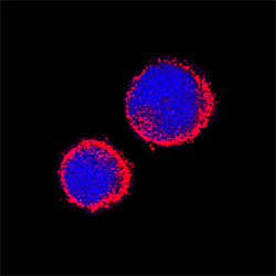
- Experimental details
- Aminopeptidase N/CD13 in Mouse Splenocytes. Aminopeptidase N/CD13 was detected in immersion fixed mouse splenocytes using Goat Anti-Mouse Aminopeptidase N/CD13 Antigen Affinity-purified Polyclonal Antibody (Catalog # AF2335) at 15 µg/mL for 3 hours at room temperature. Cells were stained using the NorthernLights™ 557-conjugated Anti-Goat IgG Secondary Antibody (red; Catalog # NL001) and counterstained with DAPI (blue). Specific staining was localized to cytoplasm. View our protocol for Fluorescent ICC Staining of Non-adherent Cells.

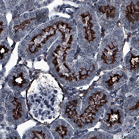
Supportive validation
- Submitted by
- R&D Systems (provider)
- Main image

- Experimental details
- Aminopeptidase N/CD13 in Mouse Kidney. Aminopeptidase N/CD13 was detected in immersion fixed paraffin-embedded sections of mouse kidney using Goat Anti-Mouse Aminopeptidase N/CD13 Antigen Affinity-purified Polyclonal Antibody (Catalog # AF2335) at 0.1 µg/mL for 1 hour at room temperature followed by incubation with the Anti-Goat IgG VisUCyte™ HRP Polymer Antibody (Catalog # VC004). Before incubation with the primary antibody, tissue was subjected to heat-induced epitope retrieval using Antigen Retrieval Reagent-Basic (Catalog # CTS013). Tissue was stained using DAB (brown) and counterstained with hematoxylin (blue). Specific staining was localized to apical membrane. View our protocol for IHC Staining with VisUCyte HRP Polymer Detection Reagents.