Explore
 Validate
 Learn
Learn Western blot
Antibody data
- Antibody Data
- Antigen structure
- References [0]
- Comments [0]
- Validations
- Western blot [2]
- Immunocytochemistry [3]
- Immunohistochemistry [5]
- Flow cytometry [1]
Submit
Validation data
Reference
Comment
Report error
- Product number
- PA5-109314 - Provider product page

- Provider
- Invitrogen Antibodies
- Product name
- CD13 Polyclonal Antibody
- Antibody type
- Polyclonal
- Antigen
- Recombinant full-length protein
- Reactivity
- Human, Mouse
- Host
- Rabbit
- Isotype
- IgG
- Vial size
- 100 µL
- Concentration
- 1 mg/mL
- Storage
- -20° C, Avoid Freeze/Thaw Cycles, store in dark
No comments: Submit comment
Supportive validation
- Submitted by
- Invitrogen Antibodies (provider)
- Main image
- Experimental details
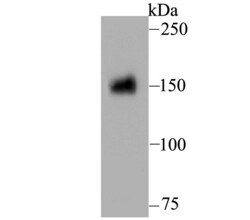
- Western blot analysis of CD13 in rat kidney tissue lysate. Samples were transferred to PVDF membrane, blocked with 5% BSA (1 hour), incubated with CD13 polyclonal antibody (Product # PA5-109314), at a dilution of 1:500 (5% BSA, 2 hours), followed by Goat Anti-Rabbit IgG-HRP (1 hour) with a dilution of 1:5000.

- Submitted by
- Invitrogen Antibodies (provider)
- Main image
- Experimental details
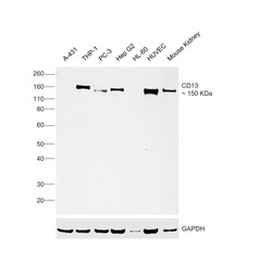
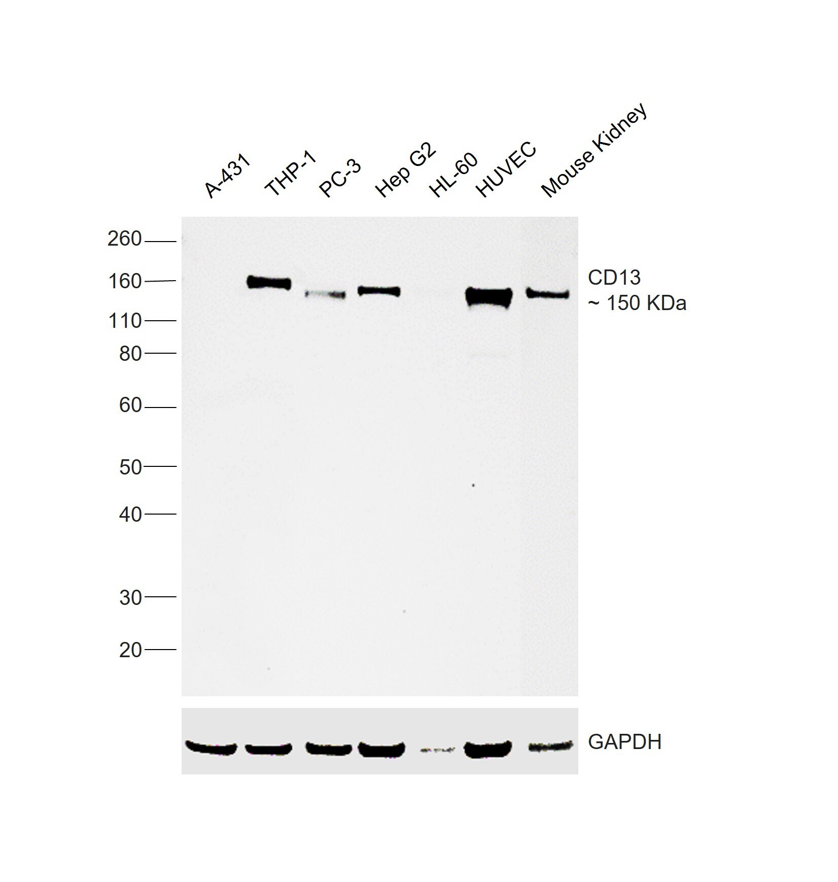
- Western blot was performed using Anti-CD13 Polyclonal Antibody (Product # PA5-109314) and a ~150 kDa band corresponding to ANPEP was observed across across cell lines tested. Membrane enriched extracts (30 µg lysate) of A-431 (Lane 1), THP-1 (Lane 2), PC-3 (Lane 3), Hep G2 (Lane 4), HL-60 (Lane 5), HUVEC (Lane 6) and Mouse Kidney (Lane 7) were electrophoresed using NuPAGE™ 4-12% Bis-Tris Protein Gel (Product # NP0322BOX), 12 well. Resolved proteins were then transferred onto a nitrocellulose membrane (Product # IB23001) by iBlot® 2 Dry Blotting System (Product # IB21001). The blot was probed with the primary antibody (1:1000 dilution) and detected by chemiluminescence with Goat anti-Rabbit IgG (H+L) Superclonal™ Recombinant Secondary Antibody, HRP (Product # A27036, 1:20,000 dilution) using the iBright™ FL1500 Imaging System (Product # A44115). Chemiluminescent detection was performed using SuperSignal™ West Pico PLUS Chemiluminescent Substrate (Product # 34580).
Supportive validation
- Submitted by
- Invitrogen Antibodies (provider)
- Main image

- Experimental details
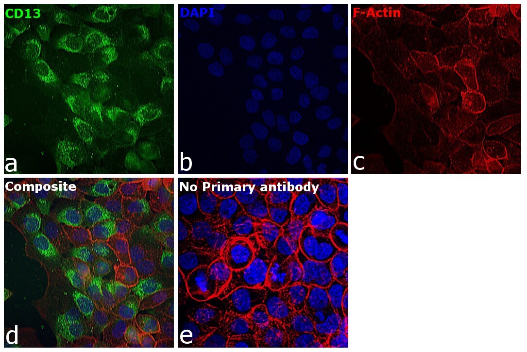
- Immunofluorescence analysis of ANPEP was performed using 70% confluent log phase A-431 cells. The cells were fixed with 4% paraformaldehyde for 10 minutes, permeabilized with 0.01% Triton™ X-100 for 15 minutes, and blocked with 2% BSA for overnight at room temperature. The cells were labeled with CD13 Polyclonal Antibody (Product # PA5-109314, 1:100 dilution) in 0.1% BSA, incubated at 4 degree celsius overnight and then labeled with Goat anti-Rabbit IgG (H+L) Superclonal™ Recombinant Secondary Antibody, Alexa Fluor® 488 conjugate (Product # A27034), (1:2000), for 45 minutes at room temperature (Panel a: Green). Nuclei (Panel b:Blue) were stained with Hoechst 33342 (Product # H1399). F-actin (Panel c: Red) was stained with Alexa Fluor™ 647 Phalloidin (Product #A22287, 1:300). Panel d represents the merged image showing cytoplasm, plasma membrane localization. Panel e represents control cells with no primary antibody to assess background. The images were captured at 40X magnification in CellInsight CX7 LZR High-Content Screening (HCS) Platform (Product # CX7A1110LZR) and externally deconvoluted (D.Sage et al./Methods 115 (2017).
- Submitted by
- Invitrogen Antibodies (provider)
- Main image
- Experimental details
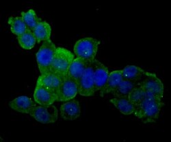
- Immunofluorescent analysis of CD13 in HT-29 cells (green). Samples were formalin fixed, permeabilized with 0.1% Triton X-100 in TBS (1 hour, room temperature) and blocked with 1% BSA (15 min, room temperature), incubated with CD13 polyclonal antibody (Product # PA5-109314) at a dilution of 1:200 (1 hour, room temperature), and followed by Alexa Fluor 488 Goat anti-Rabbit IgG and DAPI (blue) with a dilution of 1:100.

- Submitted by
- Invitrogen Antibodies (provider)
- Main image
- Experimental details
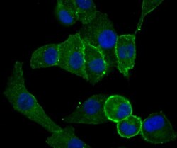
- Immunofluorescent analysis of CD13 in A549 cells (green). Samples were formalin fixed, permeabilized with 0.1% Triton X-100 in TBS (1 hour, room temperature) and blocked with 1% BSA (15 min, room temperature), incubated with CD13 polyclonal antibody (Product # PA5-109314) at a dilution of 1:500 (1 hour, room temperature), and followed by Alexa Fluor 488 Goat anti-Rabbit IgG and DAPI (blue) with a dilution of 1:100.
Supportive validation
- Submitted by
- Invitrogen Antibodies (provider)
- Main image
- Experimental details
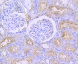
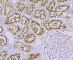
- Immunohistochemistry analysis of CD13 in paraffin-embedded rat kidney tissue. Samples were heat mediated antigen retrieval with Tris-EDTA buffer (pH 8.0-8.4, 20 minutes) and blocked in 5% BSA (30 min, room temperature), incubated with CD13 polyclonal antibody (Product # PA5-109314) at a dilution of 1:200 (30 min, room temperature), and followed by HRP conjugate, DAB and hematoxylin (mounted with DPX).
- Submitted by
- Invitrogen Antibodies (provider)
- Main image

- Experimental details
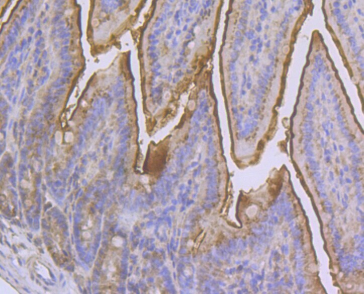
- Immunohistochemistry analysis of CD13 in paraffin-embedded mouse colon tissue. Samples were heat mediated antigen retrieval with Tris-EDTA buffer (pH 8.0-8.4, 20 minutes) and blocked in 5% BSA (30 min, room temperature), incubated with CD13 polyclonal antibody (Product # PA5-109314) at a dilution of 1:200 (30 min, room temperature), and followed by HRP conjugate, DAB and hematoxylin (mounted with DPX).
- Submitted by
- Invitrogen Antibodies (provider)
- Main image
- Experimental details
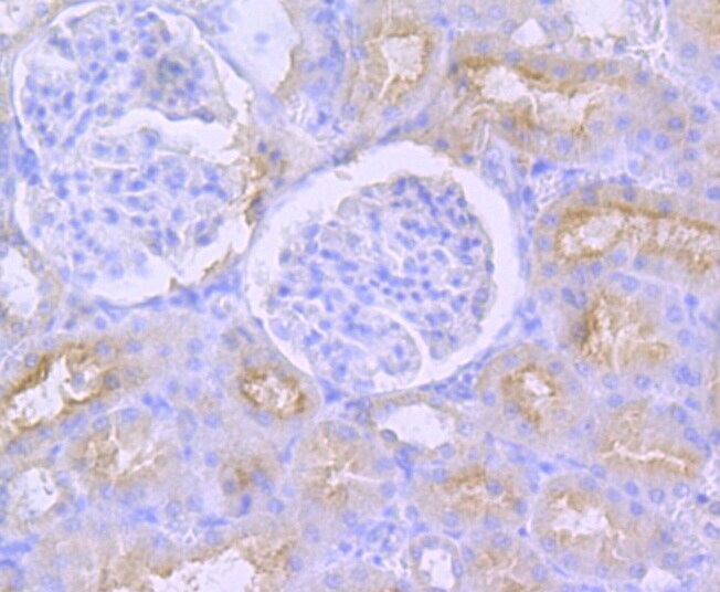
- Immunohistochemistry analysis of CD13 in paraffin-embedded human kidney tissue. Samples were heat mediated antigen retrieval with Tris-EDTA buffer (pH 8.0-8.4, 20 minutes) and blocked in 5% BSA (30 min, room temperature), incubated with CD13 polyclonal antibody (Product # PA5-109314) at a dilution of 1:200 (30 min, room temperature), and followed by HRP conjugate, DAB and hematoxylin (mounted with DPX).

- Submitted by
- Invitrogen Antibodies (provider)
- Main image
- Experimental details
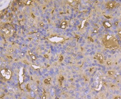
- Immunohistochemistry analysis of CD13 in paraffin-embedded human prostate cancer tissue. Samples were heat mediated antigen retrieval with Tris-EDTA buffer (pH 8.0-8.4, 20 minutes) and blocked in 5% BSA (30 min, room temperature), incubated with CD13 polyclonal antibody (Product # PA5-109314) at a dilution of 1:50 (30 min, room temperature), and followed by HRP conjugate, DAB and hematoxylin (mounted with DPX).

- Submitted by
- Invitrogen Antibodies (provider)
- Main image

- Experimental details
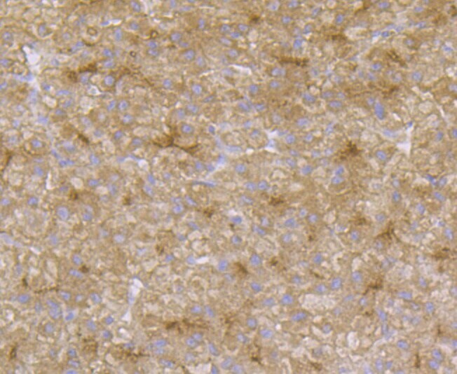
- Immunohistochemistry analysis of CD13 in paraffin-embedded human liver tissue. Samples were heat mediated antigen retrieval with Tris-EDTA buffer (pH 8.0-8.4, 20 minutes) and blocked in 5% BSA (30 min, room temperature), incubated with CD13 polyclonal antibody (Product # PA5-109314) at a dilution of 1:50 (30 min, room temperature), and followed by HRP conjugate, DAB and hematoxylin (mounted with DPX).
Supportive validation
- Submitted by
- Invitrogen Antibodies (provider)
- Main image

- Experimental details
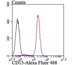
- Flow cytometry of CD13 in A549 cells (red) compared with an unlabelled control (cells without incubation with primary antibody; black). Samples were incubated with CD13 polyclonal antibody (Product # PA5-109314) at a dilution of 1:100, followed by Alexa Fluor 488-conjugated goat anti-rabbit IgG (30 min) with a dilution of 1:500.