 Explore
 Validate
 Learn
Learn Western blot
Antibody data
- Antibody Data
- Antigen structure
- References [2]
- Comments [0]
- Validations
- Western blot [1]
- ELISA [1]
- Immunohistochemistry [1]
Submit
Validation data
Reference
Comment
Report error
- Product number
- AF3345 - Provider product page

- Provider
- R&D Systems
- Product name
- Human PSGL-1/CD162 Antibody
- Antibody type
- Polyclonal
- Description
- Antigen Affinity-purified. Detects human PSGL-1/CD162 in direct ELISAs and Western blots.
- Reactivity
- Human
- Host
- Sheep
- Conjugate
- Unconjugated
- Antigen sequence
NP_002997- Isotype
- IgG
- Vial size
- 100 ug
- Concentration
- LYOPH
- Storage
- Use a manual defrost freezer and avoid repeated freeze-thaw cycles. 12 months from date of receipt, -20 to -70 °C as supplied. 1 month, 2 to 8 °C under sterile conditions after reconstitution. 6 months, -20 to -70 °C under sterile conditions after reconstitution.
Submitted references Cell surface vimentin is an attachment receptor for enterovirus 71.
Bromelain decreases neutrophil interactions with P-selectin, but not E-selectin, in vitro by proteolytic cleavage of P-selectin glycoprotein ligand-1.
Du N, Cong H, Tian H, Zhang H, Zhang W, Song L, Tien P
Journal of virology 2014 May;88(10):5816-33
Journal of virology 2014 May;88(10):5816-33
Bromelain decreases neutrophil interactions with P-selectin, but not E-selectin, in vitro by proteolytic cleavage of P-selectin glycoprotein ligand-1.
Banks JM, Herman CT, Bailey RC
PloS one 2013;8(11):e78988
PloS one 2013;8(11):e78988
No comments: Submit comment
Supportive validation
- Submitted by
- R&D Systems (provider)
- Main image
- Experimental details
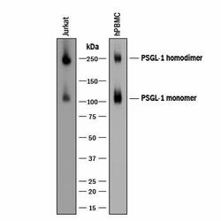
- Detection of Human PSGL-1/CD162 by Western Blot. Western blot shows lysates of Jurkat human acute T cell leukemia cell line and human peripheral blood mononuclear cells (PBMCs). PVDF membrane was probed with 0.2 µg/mL of Sheep Anti-Human PSGL-1/CD162 Antigen Affinity-purified Polyclonal Antibody (Catalog # AF3345) followed by HRP-conjugated Anti-Sheep IgG Secondary Antibody (Catalog # HAF016). Specific bands were detected for PSGL-1/CD162 homodimer at approximately 250 kDa and PSGL-1/CD162 monomer at approximately 110 kDa (as indicated). This experiment was conducted under reducing conditions and using Immunoblot Buffer Group 8.

Supportive validation
- Submitted by
- R&D Systems (provider)
- Main image

- Experimental details
- Human PSGL-1/CD162 ELISA Standard Curve. Recombinant Human PSGL-1/CD162 protein was serially diluted 2-fold and captured by Mouse Anti-Human PSGL-1/CD162 Monoclonal Antibody(Catalog # MAB9962) coated on a Clear Polystyrene Microplate (Catalog # DY990). Sheep Anti-Human PSGL-1/CD162 Antigen Affinity-purified Polyclonal Antibody (Catalog # AF3345) was biotinylated and incubated with the protein captured on the plate. Detection of the standard curve was achieved by incubating Streptavidin-HRP (Catalog # DY998) followed by Substrate Solution (Catalog # DY999) and stopping the enzymatic reaction with Stop Solution (Catalog # DY994).

Supportive validation
- Submitted by
- R&D Systems (provider)
- Main image

- Experimental details
- PSGL-1/CD162 in Human Tonsil. PSGL-1/CD162 was detected in immersion fixed paraffin-embedded sections of human tonsil using Sheep Anti-Human PSGL-1/CD162 Antigen Affinity-purified Polyclonal Antibody (Catalog # AF3345) at 3 µg/mL overnight at 4 °C. Tissue was stained using the Anti-Sheep HRP-DAB Cell & Tissue Staining Kit (brown; Catalog # CTS019) and counterstained with hematoxylin (blue). Specific staining was localized to lymphocytes. View our protocol for Chromogenic IHC Staining of Paraffin-embedded Tissue Sections.
