 Explore
 Validate
 Learn
Learn Western blot
 Immunohistochemistry
Antibody data
- Antibody Data
- Antigen structure
- References [1]
- Comments [0]
- Validations
- Western blot [4]
- Immunocytochemistry [2]
Submit
Validation data
Reference
Comment
Report error
- Product number
- PA3-16723 - Provider product page

- Provider
- Invitrogen Antibodies
- Product name
- Peripherin Polyclonal Antibody
- Antibody type
- Polyclonal
- Antigen
- Purifed from natural sources
- Reactivity
- Human, Mouse, Rat, Bovine, Porcine
- Host
- Rabbit
- Isotype
- IgG
- Vial size
- 100 µL
- Concentration
- Conc. Not Determined
- Storage
- Store at 4°C short term. For long term storage, store at -20°C, avoiding freeze/thaw cycles.
Submitted references Protective role of neuronal and lymphoid cannabinoid CB(2) receptors in neuropathic pain.
Cabañero D, Ramírez-López A, Drews E, Schmöle A, Otte DM, Wawrzczak-Bargiela A, Huerga Encabo H, Kummer S, Ferrer-Montiel A, Przewlocki R, Zimmer A, Maldonado R
eLife 2020 Jul 20;9
eLife 2020 Jul 20;9
No comments: Submit comment
Supportive validation
- Submitted by
- Invitrogen Antibodies (provider)
- Main image

- Experimental details
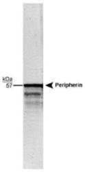
- Western Blot detection of the 57 kDa peripherin protein in whole rat brain stem homogenate using Product # PA3-16723 (1:20,000). The faint 48 kDa band is a peripherin molecule.

- Submitted by
- Invitrogen Antibodies (provider)
- Main image
- Experimental details
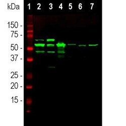
- Western blot analysis of Peripherin in tissue and cell lysates using Peripherin . Samples were incubated in Peripherin polyclonal antibody (Product # PA3-16723 using a dilution of 1:10000. Antibody in green: [1] protein standard, [2] rat spinal cord, [3] mouse spinal cord, [4] pig spinal cord, [5] cow spinal cord, [6] SH-SY5Y cells and [7] PC12 cells. The major band at ~57 kDa corresponds to the major peripherin protein isoform, while other bands presumably represent protein products of alternate transcripts of the peripherin gene.

- Submitted by
- Invitrogen Antibodies (provider)
- Main image
- Experimental details
- Western blot analysis of Peripherin in whole rat brain stem homogenate. Samples were incubated in Peripherin polyclonal antibody (Product # PA3-16723 using a dilution of 1:20,000. The faint 48 kDa band is a peripherin molecule.

- Submitted by
- Invitrogen Antibodies (provider)
- Main image

- Experimental details
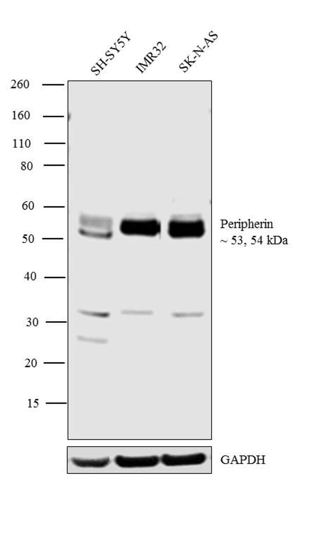
- Western blot analysis was performed on whole cell extracts of SH-SY5Y (Lane 1), IMR32 (Lane 2) and SK-N-AS (Lane 3). The blot was probed with Anti-Peripherin Polyclonal Antibody (Product # PA3-16723, 1:2500 dilution) and detected by chemiluminescence using Goat anti-Rabbit IgG (H+L) Superclonal™ Secondary Antibody, HRP conjugate (Product # A27036, 0.25 µg/ml, 1:4000 dilution). 53, 54 kDa bands corresponding to Peripherin was observed across the cell lines tested.
Supportive validation
- Submitted by
- Invitrogen Antibodies (provider)
- Main image

- Experimental details
- Immunocytochemistry analysis of Peripherin in mixed neuron/glia cultures from newborn rat brain. Samples were incubated in Peripherin polyclonal antibody (Product # PA3-16723). Antibody to peripherin (green) and chicken polyclonal antibody to phosphorylated NF-H (red channel). A class of large neurons, like the one at the top right of this image, contain peripherin, while the majority of neurons and their processes contain NF-H and not peripherin. The blue channel reveals the localization of DNA.

- Submitted by
- Invitrogen Antibodies (provider)
- Main image

- Experimental details
- Immunocytochemistry analysis of Peripherin in rat pheochromocytoma PC12 cell line. Samples were incubated in Peripherin polyclonal antibody (Product # PA3-16723) using a dilution of 1:2000. Periphren antibody (Green). Hoechst staining of nuclear DNA (Blue). Peripherin, one of the Class III family of intermediate filament subunit proteins, is a major component of the PC12 cell forming a perinuclear cap, with some filaments visible in the cytoplasm.
