Explore
 Validate
 Learn
Learn Immunocytochemistry
 Flow cytometry
Antibody data
- Antibody Data
- Antigen structure
- References [3]
- Comments [0]
- Validations
- Immunocytochemistry [1]
- Other assay [1]
Submit
Validation data
Reference
Comment
Report error
- Product number
- MAB6321-100 - Provider product page

- Provider
- R&D Systems
- Product name
- Human CD40/TNFRSF5 Antibody
- Antibody type
- Monoclonal
- Description
- Protein A or G purified from hybridoma culture supernatant. Detects human CD40/TNFRSF5 in direct ELISAs and Western blots. In direct ELISAs, does not cross-react with recombinant human (rh) 4-1BB, rhCD27, rhCD30, recombinant mouse CD40, rhDR3, rhDR6, rhEDAR, rhFas, rhGITR, rhHVEM, rhLTR beta , rhNGF R, rhOPG, rhRANK, rhTAJ, rhTNF RI, or rhTNF RII.
- Reactivity
- Human
- Host
- Mouse
- Conjugate
- Unconjugated
- Antigen sequence
P25942- Isotype
- IgG
- Antibody clone number
- 82111
- Vial size
- 100 ug
- Storage
- Use a manual defrost freezer and avoid repeated freeze-thaw cycles. 12 months from date of receipt, -20 to -70 °C as supplied. 1 month, 2 to 8 °C under sterile conditions after reconstitution. 6 months, -20 to -70 °C under sterile conditions after reconstitution.
Submitted references Deficient EBV-specific B- and T-cell response in patients with chronic fatigue syndrome.
Autocrine IL-10 promotes human B-cell differentiation into IgM- or IgG-secreting plasmablasts.
Fibrosis in human adipose tissue: composition, distribution, and link with lipid metabolism and fat mass loss.
Loebel M, Strohschein K, Giannini C, Koelsch U, Bauer S, Doebis C, Thomas S, Unterwalder N, von Baehr V, Reinke P, Knops M, Hanitsch LG, Meisel C, Volk HD, Scheibenbogen C
PloS one 2014;9(1):e85387
PloS one 2014;9(1):e85387
Autocrine IL-10 promotes human B-cell differentiation into IgM- or IgG-secreting plasmablasts.
Heine G, Drozdenko G, Grün JR, Chang HD, Radbruch A, Worm M
European journal of immunology 2014 Jun;44(6):1615-21
European journal of immunology 2014 Jun;44(6):1615-21
Fibrosis in human adipose tissue: composition, distribution, and link with lipid metabolism and fat mass loss.
Divoux A, Tordjman J, Lacasa D, Veyrie N, Hugol D, Aissat A, Basdevant A, Guerre-Millo M, Poitou C, Zucker JD, Bedossa P, Clément K
Diabetes 2010 Nov;59(11):2817-25
Diabetes 2010 Nov;59(11):2817-25
No comments: Submit comment
Supportive validation
- Submitted by
- R&D Systems (provider)
- Main image

- Experimental details
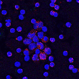
- CD40/TNFRSF5 in Human PBMCs. CD40/TNFRSF5 was detected in immersion fixed human peripheral blood mononuclear cells (PBMCs) using Mouse Anti-Human CD40/TNFRSF5 Monoclonal Antibody (Catalog # MAB6321) at 10 µg/mL for 3 hours at room temperature. Cells were stained using the NorthernLights™ 557-conjugated Anti-Mouse IgG Secondary Antibody (red; Catalog # NL007) and counterstained with DAPI (blue). Specific staining was localized to cell surfaces and cytoplasm. View our protocol for Fluorescent ICC Staining of Non-adherent Cells.
Supportive validation
- Submitted by
- R&D Systems (provider)
- Main image
- Experimental details
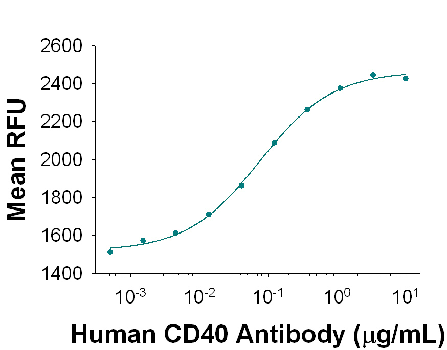
- Human CD40/TNFRSF5 Antibody Stimulates Cell Proliferation in Human B Cells. Mouse Anti-Human CD40/TNFRSF5 Monoclonal Antibody (Catalog # MAB6321) stimulates human B cell proliferation in the presence of Recombinant Human IL-4 (Catalog # 204-IL) in a dose-dependent manner, as measured by Resazurin (Catalog # AR002). The ED50 for this effect is typically 0.035-0.175 μg/mL