Antibody data
- Antibody Data
- Antigen structure
- References [0]
- Comments [0]
- Validations
- Western blot [1]
- ELISA [1]
- Flow cytometry [1]
Submit
Validation data
Reference
Comment
Report error
- Product number
- AM33033PU-N - Provider product page

- Provider
- Acris Antibodies GmbH
- Product name
- anti CD50 / ICAM3
- Antibody type
- Monoclonal
- Antigen
- Derived by fusion of mouse Ag8.653 cells with spleen cells from a BALB/c mouse immunized with an ICAM-3/HEK transfectant.
- Reactivity
- Human
- Host
- Mouse
- Isotype
- IgG
- Antibody clone number
- MA4
- Vial size
- 0.1 mg
- Concentration
- 1.0 mg/ml
No comments: Submit comment
Supportive validation
- Submitted by
- Acris Antibodies GmbH (provider)
- Main image

- Experimental details
- Purified ICAM-3-Fc was separated by using PAGE and transferred via electroblotting to nitrocellulose membrane. The membrane was probed with MA4 at a dilution of 1/200 (using TBST â TBS+0.05% tween 20) which equates to a concentration of 5 µg/ml and mAb binding detected by anti-mouse-HRP (1/1000). This single band on western blot analysis is consistent with the published reports using this recombinant ICAM-3-Fc which contained only domains 1 and 2 of ICAM-3 fused to human Fc. The band shown is approximately 80kDa.
Supportive validation
- Submitted by
- Acris Antibodies GmbH (provider)
- Main image

- Experimental details
- Purified ICAM-3-Fc or CD14-Fc was captured to an ELISA plate and probed with the indicated dilution of mAb MA4. The binding of mAb was detected anti-Mouse-HRP (1/1000) and OPD detection system. Data shown are mean ± SD of a representative experiment. MA4 shows clear specificity for ICAM-3.
Supportive validation
- Submitted by
- Acris Antibodies GmbH (provider)
- Main image

- Experimental details
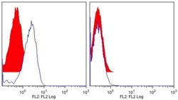
- Mutu (human B cell line) that were wild type (left panel) or ICAM-3-deficient (right panel) were stained with mAb MA4 (blue line) or an isotype control (solid red). Per tube, 2 x 10⁵ cells were stained with 75 µl of mAb at the indicated concentrations. Following 30 min incubation at 4°C, unbound Ab was removed by washing and bound mAb detected by staining with goat anti-mouse-PE for 30 min at 4°C. Flow cytometric histograms of 5000 events per sample are shown.
 Explore
 Validate
 Learn
Learn Western blot
 Immunocytochemistry