Explore
 Validate
 Learn
Learn Western blot
Antibody data
- Antibody Data
- Antigen structure
- References [9]
- Comments [0]
- Validations
- Western blot [1]
- Immunocytochemistry [1]
- Immunohistochemistry [3]
- Other assay [1]
Submit
Validation data
Reference
Comment
Report error
- Product number
- MA1-24654 - Provider product page

- Provider
- Invitrogen Antibodies
- Product name
- Tyrosine Hydroxylase Monoclonal Antibody (185)
- Antibody type
- Monoclonal
- Antigen
- Recombinant full-length protein
- Description
- Store product as a concentrated solution. Centrifuge briefly prior to opening the vial.
- Reactivity
- Human, Mouse, Rat
- Host
- Mouse
- Isotype
- IgG
- Antibody clone number
- 185
- Vial size
- 50 µL
- Concentration
- 0.24 mg/mL
- Storage
- Store at 4°C short term. For long term storage, store at -20°C, avoiding freeze/thaw cycles.
Submitted references Somatostatin Neurons in the Mouse Pontine Nucleus Activate GABA(A) Receptor Mediated Synaptic Currents in Locus Coeruleus Neurons.
Lack of Mucosal Cholinergic Innervation Is Associated With Increased Risk of Enterocolitis in Hirschsprung's Disease.
Restoration of Noradrenergic Function in Parkinson's Disease Model Mice.
Membrane Trafficking Protein CDP138 Regulates Fat Browning and Insulin Sensitivity through Controlling Catecholamine Release.
An autopsy case of microencephaly, bizarre putaminal lesion, and cerebellar atrophy with heart and liver diseases.
Melatonin alterations and brain acetylcholine lesions in sleep disorders in Cockayne syndrome.
Lesions of acetylcholine neurons in refractory epilepsy.
Intestinal epithelial stem/progenitor cells are controlled by mucosal afferent nerves.
An autopsy case presenting repetitive hypoglycemia and unique cortical dysplasia.
Garcia DuBar S, Cosio D, Korthas H, Van Batavia JP, Zderic SA, Sahibzada N, Valentino RJ, Vicini S
Frontiers in synaptic neuroscience 2021;13:754786
Frontiers in synaptic neuroscience 2021;13:754786
Lack of Mucosal Cholinergic Innervation Is Associated With Increased Risk of Enterocolitis in Hirschsprung's Disease.
Keck S, Galati-Fournier V, Kym U, Moesch M, Usemann J, Müller I, Subotic U, Tharakan SJ, Krebs T, Stathopoulos E, Schmittenbecher P, Cholewa D, Romero P, Reingruber B, Bruder E, Group NS, Holland-Cunz S
Cellular and molecular gastroenterology and hepatology 2021;12(2):507-545
Cellular and molecular gastroenterology and hepatology 2021;12(2):507-545
Restoration of Noradrenergic Function in Parkinson's Disease Model Mice.
Cui K, Yang F, Tufan T, Raza MU, Zhan Y, Fan Y, Zeng F, Brown RW, Price JB, Jones TC, Miller GW, Zhu MY
ASN neuro 2021 Jan-Dec;13:17590914211009730
ASN neuro 2021 Jan-Dec;13:17590914211009730
Membrane Trafficking Protein CDP138 Regulates Fat Browning and Insulin Sensitivity through Controlling Catecholamine Release.
Zhou QL, Song Y, Huang CH, Huang JY, Gong Z, Liao Z, Sharma AG, Greene L, Deng JZ, Rigor MC, Xie X, Qi S, Ayala JE, Jiang ZY
Molecular and cellular biology 2018 Apr 15;38(8)
Molecular and cellular biology 2018 Apr 15;38(8)
An autopsy case of microencephaly, bizarre putaminal lesion, and cerebellar atrophy with heart and liver diseases.
Okoshi Y, Hayashi M, Kanda S, Yamamoto T
Brain & development 2014 Sep;36(8):707-10
Brain & development 2014 Sep;36(8):707-10
Melatonin alterations and brain acetylcholine lesions in sleep disorders in Cockayne syndrome.
Okoshi Y, Tanuma N, Miyata R, Hayashi M
Brain & development 2014 Nov;36(10):907-13
Brain & development 2014 Nov;36(10):907-13
Lesions of acetylcholine neurons in refractory epilepsy.
Hayashi M, Nakajima K, Miyata R, Tanuma N, Kodama T
ISRN neurology 2012;2012:404263
ISRN neurology 2012;2012:404263
Intestinal epithelial stem/progenitor cells are controlled by mucosal afferent nerves.
Lundgren O, Jodal M, Jansson M, Ryberg AT, Svensson L
PloS one 2011 Feb 9;6(2):e16295
PloS one 2011 Feb 9;6(2):e16295
An autopsy case presenting repetitive hypoglycemia and unique cortical dysplasia.
Hayashi M, Hachiya Y, Arai N
Brain & development 2010 Apr;32(4):289-92
Brain & development 2010 Apr;32(4):289-92
No comments: Submit comment
Supportive validation
- Submitted by
- Invitrogen Antibodies (provider)
- Main image
- Experimental details
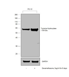
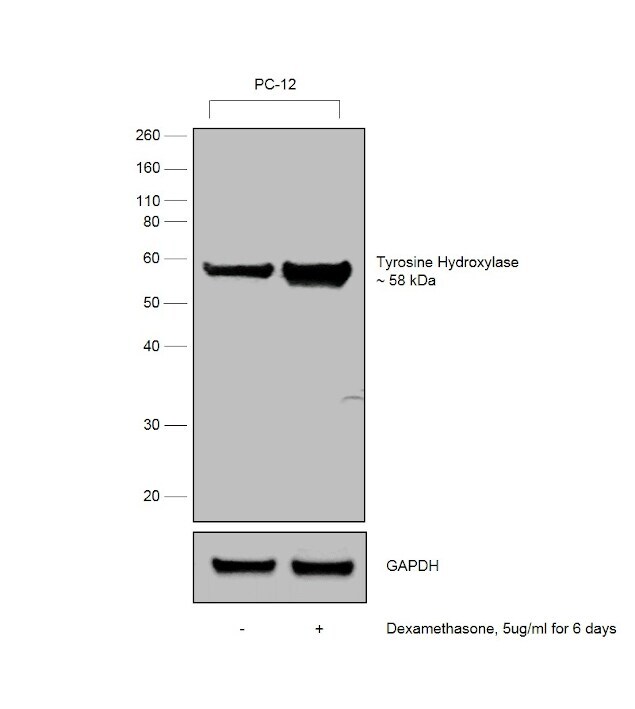
- Western blot was performed using Anti-Tyrosine Hydroxylase Monoclonal Antibody (185) (Product # MA1-24654) and 58 kDa band corresponding to Tyrosine Hydroxylase was observed and enhanced upon Dexamethasone treatment in PC-12 cell line. Dexamethasone, a synthetic glucocorticoid, is known to increase the expression of Tyrosine Hydroxylase in neuronal cells. Whole cell extracts (30 µg lysate) of PC-12 (Lane1) and PC-12 treated with dexamethasone (5ug/ml for 6 days) (Lane2) were electrophoresed using Novex® NuPAGE® 4-12% Bis-Tris gel (Product # NP0342BOX). Resolved proteins were then transferred onto a nitrocellulose membrane (Product # IB23001) by iBlot® 2 Dry Blotting System (Product # IB21001). The blot was probed with the primary antibody (1:500 dilution) and detected by Goat anti-Mouse IgG (H+L) Superclonal™ Recombinant Secondary Antibody, HRP (Product # A28177) , 1:4000 dilution) using the iBright FL 1000 (Product # A32752). Chemiluminescent detection was performed using Novex® ECL Chemiluminescent Substrate Reagent Kit (Product # WP20005).
Supportive validation
- Submitted by
- Invitrogen Antibodies (provider)
- Main image
- Experimental details
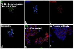
- Immunofluorescence analysis of Tyrosine Hydroxylase using of Tyrosine Hydroxylase using Tyrosine Hydroxylase Monoclonal Antibody (185) (Product # MA1-24654) was performed using 70% confluent log phase PC-12 cells treated with 5 µg/mL dexamethasone for 6 days. The cells were fixed with 4% Paraformaldehyde for 10 minutes, permeabilized with 0.1% Triton™ X-100 for 10 minutes, and blocked with 2% BSA for 10 minutes at room temperature. The cells were labeled with Tyrosine Hydroxylase Monoclonal Antibody (185) (Product # MA1-24654) at 1:50 µg/mL in 0.1% BSA, incubated at 4 degree Celsius overnight and then labeled with Goat anti-Mouse IgG (H+L), Superclonal™ Recombinant Secondary Antibody, Alexa Fluor 488 (Product # A28175), (1:2000 dilution) for 45 minutes at room temperature (Panel a: Green). Nuclei (Panel b: Blue) were stained with SlowFade® Gold Antifade Mountant with DAPI (Product # S36938). F-actin (Panel c: Red) was stained with Rhodamine Phalloidin (Product # R415, 1:300). Panel d represents the merged image showing Cytoplasmic localization. Panel e represents untreated PC-12 cells having low expression of TH. Panel f represents control cells with no primary antibody to assess background. The images were captured at 60X magnification.

Supportive validation
- Submitted by
- Invitrogen Antibodies (provider)
- Main image
- Experimental details
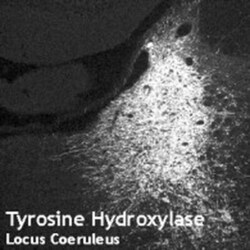
- Immunohistochemistry analysis of locus coeruleus using Tyrosine Hydroxylase Monoclonal Antibody (185) (Product # MA1-24654).

- Submitted by
- Invitrogen Antibodies (provider)
- Main image
- Experimental details
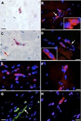
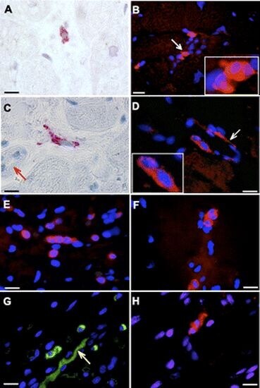
- Immunohistochemistry analysis of human heart tissue (labeling of intrinsic cardiac adrenergic (ICA) cells) using Tyrosine Hydroxylase Monoclonal Antibody (185) (Product # MA1-24654).
- Submitted by
- Invitrogen Antibodies (provider)
- Main image
- Experimental details
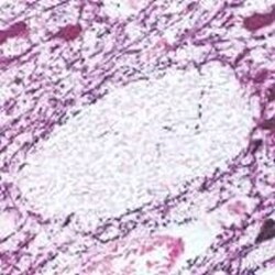
- Immunohistochemical (Paraffin) analysis of human mid-brain tissue using (Product # MA1-24654) Tyrosine Hydroxylase Monoclonal Antibody (185).

Supportive validation
- Submitted by
- Invitrogen Antibodies (provider)
- Main image
- Experimental details
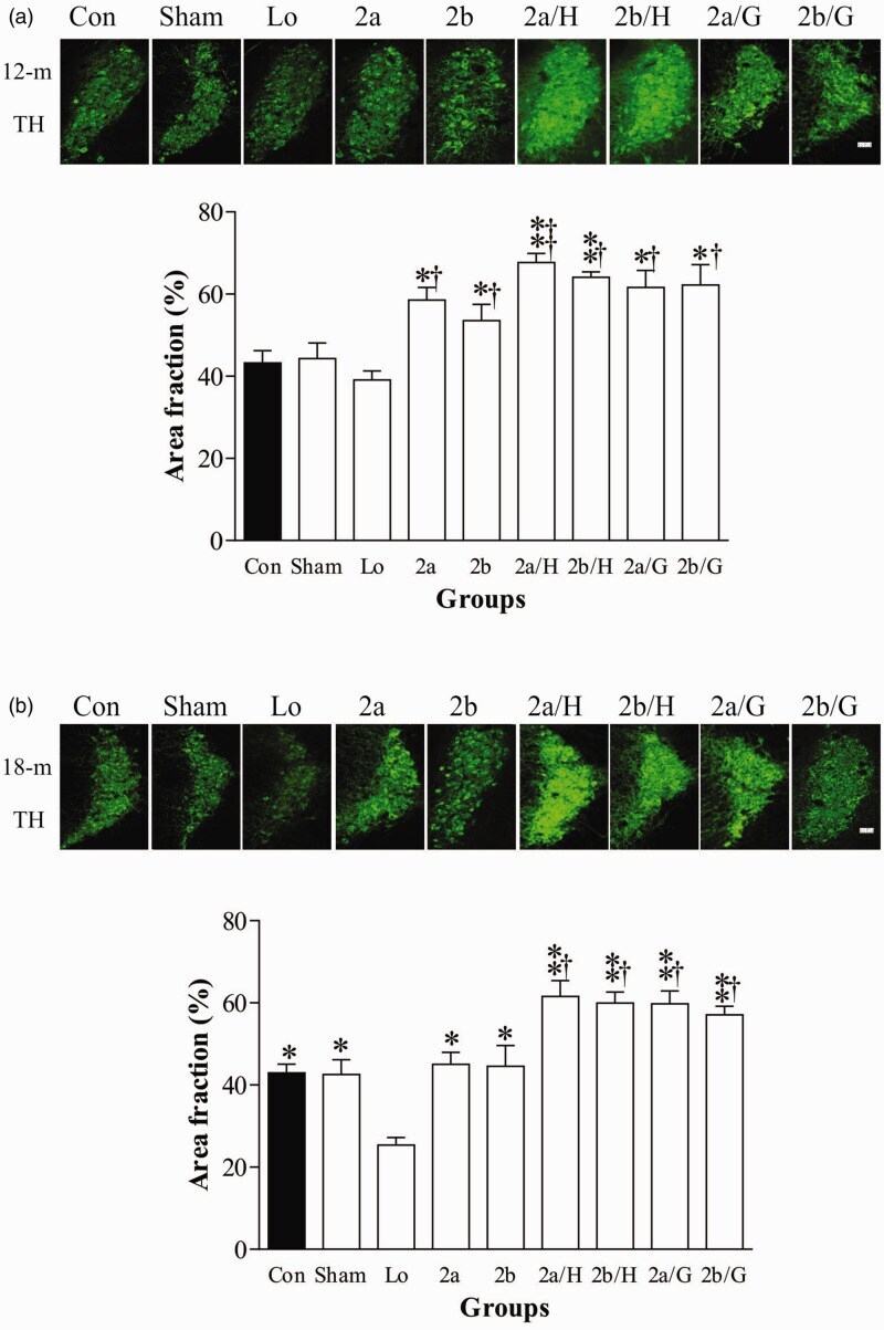
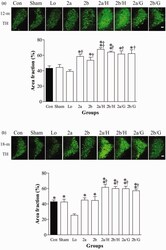
- Figure 4. Effects of microinjection on TH proteins in the LC. after microinjection of lentiviral expression cassettes (N = 6) measured by immunoflurescence in mice at age of 12 month-old (A) and 18 month-old (N = 5) (B). Upper panels in A and B are representative micrographs of TH immunofluorescence. Lower panels in A and B: quantitative analysis of TH immunofluorescence. *p < 0.05, **p < 0.01, compared to the Lo mice + p < 0.05, compared to the control. See legends of Figures 1 and 2 for abbreviations. Scale bar: 25 um for all images.