Antibody data
- Antibody Data
- Antigen structure
- References [4]
- Comments [0]
- Validations
- Immunocytochemistry [1]
- Immunohistochemistry [1]
Submit
Validation data
Reference
Comment
Report error
- Product number
- GTX10372 - Provider product page

- Provider
- GeneTex
- Proper citation
- GeneTex Cat#GTX10372, RRID:AB_373127
- Product name
- Tyrosine Hydroxylase antibody [185]
- Antibody type
- Monoclonal
- Reactivity
- Human, Mouse, Rat
- Host
- Mouse
Submitted references Two Subpopulations of Noradrenergic Neurons in the Locus Coeruleus Complex Distinguished by Expression of the Dorsal Neural Tube Marker Pax7.
Critical role of TRPC1 in thyroid hormone-dependent dopaminergic neuron development.
Uncovering diversity in the development of central noradrenergic neurons and their efferents.
Expanding the power of recombinase-based labeling to uncover cellular diversity.
Plummer NW, Scappini EL, Smith KG, Tucker CJ, Jensen P
Frontiers in neuroanatomy 2017;11:60
Frontiers in neuroanatomy 2017;11:60
Critical role of TRPC1 in thyroid hormone-dependent dopaminergic neuron development.
Chen C, Ma Q, Deng P, Yang J, Yang L, Lin M, Yu Z, Zhou Z
Biochimica et biophysica acta. Molecular cell research 2017 Oct;1864(10):1900-1912
Biochimica et biophysica acta. Molecular cell research 2017 Oct;1864(10):1900-1912
Uncovering diversity in the development of central noradrenergic neurons and their efferents.
Robertson SD, Plummer NW, Jensen P
Brain research 2016 Jun 15;1641(Pt B):234-44
Brain research 2016 Jun 15;1641(Pt B):234-44
Expanding the power of recombinase-based labeling to uncover cellular diversity.
Plummer NW, Evsyukova IY, Robertson SD, de Marchena J, Tucker CJ, Jensen P
Development (Cambridge, England) 2015 Dec 15;142(24):4385-93
Development (Cambridge, England) 2015 Dec 15;142(24):4385-93
No comments: Submit comment
Supportive validation
- Submitted by
- GeneTex (provider)
- Main image

- Experimental details
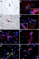
- Immunoperoxidase (A and C) and immunofluorescent (B, D¡VH) labeling of intrinsic cardiac adrenergic (ICA) cells in human hearts are shown. ICA cells expressing tyrosine hydroxylase (TH) immunoreactivity (red) are distributed diffusely throughout the left ventricular (LV) myocardium. Perivascular location is a frequent feature of ICA cells. C, arrow: terminal arteriole. E: abundant ICA cells in the smooth muscle layers of epicardial circumflex coronary artery. TH-expressing sympathetic nerve fibers (D and G, arrows) can occasionally be seen in the field. B and D, insets: magnified ICA cell images (arrows). TH immunoreactivity (green) was identified in ICA cells and sympathetic nerve fibers in the sinoatrial nodal tissue (G). ICA cells are seen in transplanted human LV tissue (H). Scale bars = 10 ?m, except in B (20 ?m)
Supportive validation
- Submitted by
- GeneTex (provider)
- Main image

- Experimental details
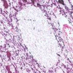
- TH staining of human mid-brain. Note cytoplasmic staining of catecholaminergic cells and their processes. Paraffin section (Peroxidase substrate: nickel DAB, Counterstain: eosin).
 Explore
 Validate
 Learn
Learn Western blot
 Immunocytochemistry