Antibody data
- Antibody Data
- Antigen structure
- References [1]
- Comments [0]
- Validations
- Western blot [1]
- Immunocytochemistry [1]
- Immunohistochemistry [1]
- Flow cytometry [4]
Submit
Validation data
Reference
Comment
Report error
- Product number
- NBP2-42212 - Provider product page

- Provider
- Novus Biologicals
- Product name
- Mouse Monoclonal Tyrosine Hydroxylase Antibody
- Antibody type
- Monoclonal
- Description
- Protein G purified.
- Reactivity
- Human, Rat
- Host
- Mouse
- Isotype
- IgG
- Vial size
- 0.1 mg
- Concentration
- 1.0 mg/ml
- Storage
- Store at -20C. Avoid freeze-thaw cycles.
Submitted references Rescue of Pink1 Deficiency by Stress-Dependent Activation of Autophagy.
Zhang Y, Nguyen DT, Olzomer EM, Poon GP, Cole NJ, Puvanendran A, Phillips BR, Hesselson D
Cell chemical biology 2017 Apr 20;24(4):471-480.e4
Cell chemical biology 2017 Apr 20;24(4):471-480.e4
No comments: Submit comment
Supportive validation
- Submitted by
- Novus Biologicals (provider)
- Main image
- Experimental details
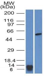
- Western Blot: Tyrosine Hydroxylase Antibody (2D3.1G7) [NBP2-42212] - WB analysis of a partial - recombinant human Tyrosine Hydroxylase protein and a lysate of HEK293 cells using 3 ug/ml concentration of Tyrosine Hydroxylase antibody (clone 2D3.1G7). The antibody detected ~11 kDa and ~58 kDa specific bands representing the recombinant and the endogenous TH proteins respectively.
Supportive validation
- Submitted by
- Novus Biologicals (provider)
- Main image
- Experimental details
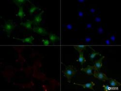
- Immunocytochemistry/Immunofluorescence: Tyrosine Hydroxylase Antibody (2D3.1G7) [NBP2-42212] - PC-12 cells were fixed for 10 minutes using 10% formalin and then permeabilized for 5 minutes using 1X TBS + 0.5% Triton-X100. The cells were incubated with Tyrosine Hydroxylase (2D3.1G7) at a 1:100 dilution overnight at 4 degrees Celsius and detected with Dylight 488 (Green). Actin was detected with Phalloidin 568 (Red) at a 1:200 dilution. Nuclei were detected with DAPI (Blue). Cells were imaged using a 40X objective.
Supportive validation
- Submitted by
- Novus Biologicals (provider)
- Main image
- Experimental details
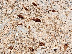
- Immunohistochemistry: Tyrosine Hydroxylase Antibody (2D3.1G7) [NBP2-42212] - IHC analysis of a formalin fixed paraffin embedded tissue section of human brain/substantia nigra using Tyrosine Hydroxylase antibody (clone 2D3.1G7) at 15 ug/ml concentration. This representative photomicrograph shows strong immunostaining of tyrosine hydroxylase/TH in TH-positive neurons.
Supportive validation
- Submitted by
- Novus Biologicals (provider)
- Main image
- Experimental details
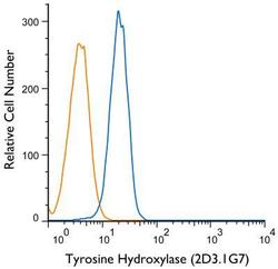
- Flow (Intracellular): Tyrosine Hydroxylase Antibody (2D3.1G7) [NBP2-42212] - An intracellular stain was performed on SH-SY5Y cells with Tyrosine Hydroxylase (2D3.1G7) antibody NBP2-42212 (blue) and a matched isotype control NBP1-97005 (orange). Cells were fixed with 4% PFA and then permeablized with 0.1% saponin. Cells were incubated in an antibody dilution of 2.5 ug/mL for 30 minutes at room temperature, followed by mouse F(ab)2 IgG (H+L) PE-conjugated Antibody (F0102B, R&D systems).
- Submitted by
- Novus Biologicals (provider)
- Main image
- Experimental details
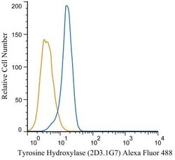
- Flow Cytometry: Tyrosine Hydroxylase Antibody (2D3.1G7) [NBP2-42212] - An intracellular stain was performed on SH-SY5Y cells with Tyrosine Hydroxylase (2D3.1G7) antibody NBP2-42212AF488 (blue) and a matched isotype control NB600-986AF488 (orange). Cells were fixed with 4% PFA and then permeablized with 0.1% saponin. Cells were incubated in an antibody dilution of 5 ug/mL for 30 minutes at room temperature. Both antibodies were conjugated to Alexa Fluor 488.
- Submitted by
- Novus Biologicals (provider)
- Main image
- Experimental details
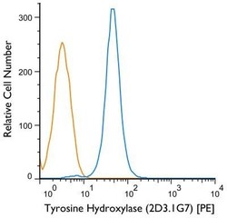
- Flow Cytometry: Tyrosine Hydroxylase Antibody (2D3.1G7) [NBP2-42212] - Using the PE direct conjugate An intracellular stain was performed on SH-SY5Y cells with Tyrosine Hydroxylase (2D3.1G7) antibody NBP2-42212PE (blue) and a matched isotype control NBP1-97005PE (orange). Cells were fixed with 4% PFA and then permeablized with 0.1% saponin. Cells were incubated in an antibody dilution of 1 ug/mL for 30 minutes at room temperature. Both antibodies were conjugated to phycoerythrin.
- Submitted by
- Novus Biologicals (provider)
- Main image
- Experimental details
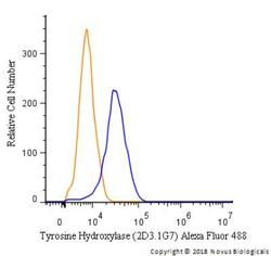
- Flow Cytometry: Tyrosine Hydroxylase Antibody (2D3.1G7) [NBP2-42212] - An intracellular stain was performed on PC12 cells with Tyrosine Hydroxylase [2D3.1G7] Antibody NBP2-42212AF488 (blue) and a matched isotype control (orange). Cells were fixed with 4% PFA and then permeabilized with 0.1% saponin. Cells were incubated in an antibody dilution of 5 ug/mL for 30 minutes at room temperature. Both antibodies were conjugated to Alexa Fluor 488.
 Explore
 Validate
 Learn
Learn Western blot