 Explore
 Validate
 Learn
Learn Western blot
Antibody data
- Antibody Data
- Antigen structure
- References [4]
- Comments [0]
- Validations
- Western blot [2]
- Immunocytochemistry [1]
- Immunohistochemistry [1]
Submit
Validation data
Reference
Comment
Report error
- Product number
- MAB7566 - Provider product page

- Provider
- R&D Systems
- Product name
- Human/Mouse Tyrosine Hydroxylase Antibody
- Antibody type
- Monoclonal
- Description
- Protein A or G purified from hybridoma culture supernatant. Detects human Tyrosine Hydroxylase in direct ELISAs and human, mouse and rat Tyrosine Hydroxylase in Western blots. In direct ELISAs, no cross-reactivity with recombinant human Tryptophan Hydroxylase-1 is observed.
- Reactivity
- Human, Mouse
- Host
- Mouse
- Conjugate
- Unconjugated
- Antigen sequence
P07101- Isotype
- IgG
- Antibody clone number
- 779427
- Vial size
- 100 ug
- Concentration
- LYOPH
- Storage
- Use a manual defrost freezer and avoid repeated freeze-thaw cycles. 12 months from date of receipt, -20 to -70 °C as supplied. 1 month, 2 to 8 °C under sterile conditions after reconstitution. 6 months, -20 to -70 °C under sterile conditions after reconstitution.
Submitted references Upregulation of neuronal astrocyte elevated gene-1 protects nigral dopaminergic neurons in vivo.
A single cell high content assay detects mitochondrial dysfunction in iPSC-derived neurons with mutations in SNCA.
Single-Factor SOX2 Mediates Direct Neural Reprogramming of Human Mesenchymal Stem Cells via Transfection of In Vitro Transcribed mRNA.
27-Hydroxycholesterol increases α-synuclein protein levels through proteasomal inhibition in human dopaminergic neurons.
Leem E, Kim HJ, Choi M, Kim S, Oh YS, Lee KJ, Choe YS, Um JY, Shin WH, Jeong JY, Jin BK, Kim DW, McLean C, Fisher PB, Kholodilov N, Ahn KS, Lee JM, Jung UJ, Lee SG, Kim SR
Cell death & disease 2018 May 1;9(5):449
Cell death & disease 2018 May 1;9(5):449
A single cell high content assay detects mitochondrial dysfunction in iPSC-derived neurons with mutations in SNCA.
Little D, Luft C, Mosaku O, Lorvellec M, Yao Z, Paillusson S, Kriston-Vizi J, Gandhi S, Abramov AY, Ketteler R, Devine MJ, Gissen P
Scientific reports 2018 Jun 13;8(1):9033
Scientific reports 2018 Jun 13;8(1):9033
Single-Factor SOX2 Mediates Direct Neural Reprogramming of Human Mesenchymal Stem Cells via Transfection of In Vitro Transcribed mRNA.
Kim BE, Choi SW, Shin JH, Kim JJ, Kang I, Lee BC, Lee JY, Kook MG, Kang KS
Cell transplantation 2018 Jul;27(7):1154-1167
Cell transplantation 2018 Jul;27(7):1154-1167
27-Hydroxycholesterol increases α-synuclein protein levels through proteasomal inhibition in human dopaminergic neurons.
Schommer J, Marwarha G, Schommer T, Flick T, Lund J, Ghribi O
BMC neuroscience 2018 Apr 3;19(1):17
BMC neuroscience 2018 Apr 3;19(1):17
No comments: Submit comment
Supportive validation
- Submitted by
- R&D Systems (provider)
- Main image
- Experimental details
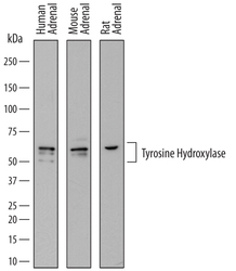
- Detection of Human, Mouse, and Rat Tyrosine Hydroxylase by Western Blot. Western blot shows lysates of human adrenal gland tissue, mouse adrenal gland tissue, and rat adrenal gland tissue. PVDF membrane was probed with 0.25 µg/mL of Mouse Anti-Human/Mouse Tyrosine Hydroxylase Monoclonal Antibody (Catalog # MAB7566) followed by HRP-conjugated Anti-Mouse IgG Secondary Antibody (Catalog # HAF018). Specific bands were detected for Tyrosine Hydroxylase at approximately 50-60 kDa (as indicated). This experiment was conducted under reducing conditions and using Immunoblot Buffer Group 1.

- Submitted by
- R&D Systems (provider)
- Main image

- Experimental details
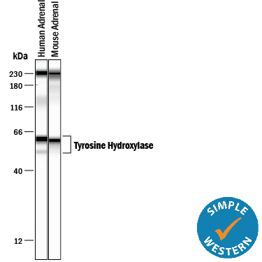
- Detection of Human and Mouse Tyrosine Hydroxylase by Simple WesternTM. Simple Western lane view shows lysates of human adrenal gland tissue and mouse adrenal gland tissue, loaded at 0.2 mg/mL. Specific bands were detected for Tyrosine Hydroxylase at approximately 52-61 kDa (as indicated) using 5 µg/mL of Mouse Anti-Human/Mouse Tyrosine Hydroxylase Monoclonal Antibody (Catalog # MAB7566). This experiment was conducted under reducing conditions and using the 12-230 kDa separation system. Non-specific interaction with the 230 kDa Simple Western standard may be seen with this antibody.
Supportive validation
- Submitted by
- R&D Systems (provider)
- Main image

- Experimental details
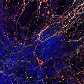
- Tyrosine Hydroxylase in Mouse Dopaminergic Neurons. Tyrosine Hydroxylase was detected in immersion fixed mouse embryonic stem cells differentiated into dopaminergic neurons using Mouse Anti-Human/Mouse Tyrosine Hydroxylase Monoclonal Antibody (Catalog # MAB7566) at 10 µg/mL for 3 hours at room temperature. Cells were stained using the Northern-Lights™ 557-conjugated Anti-Mouse IgG Secondary Antibody (red; Catalog # NL007). Cells were double stained using the Northern-Lights™ 637-conjugated Mouse Anti-Neuron-specific beta-III Tubulin Monoclonal Antibody (white; Catalog # NL1195V). Cells were counterstained with DAPI (blue). Specific staining of Tyrosine Hydroxylase was localized to cytoplasm of dopaminergic neurons. View our protocol for Fluorescent ICC Staining of Cells on Coverslips.
Supportive validation
- Submitted by
- R&D Systems (provider)
- Main image
- Experimental details
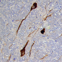
- Tyrosine Hydroxylase in Human Brain. Tyrosine Hydroxylase was detected in immersion fixed paraffin-embedded sections of human brain (medulla) using Mouse Anti-Human/Mouse Tyrosine Hydroxylase Monoclonal Antibody (Catalog # MAB7566) at 25 µg/mL overnight at 4 °C. Tissue was stained using the Anti-Mouse HRP-DAB Cell & Tissue Staining Kit (brown; Catalog # CTS002) and counterstained with hematoxylin (blue). Specific staining was localized to neurons. View our protocol for Chromogenic IHC Staining of Paraffin-embedded Tissue Sections.
