 Explore
 Validate
 Learn
Learn Western blot
 ELISA
ELISAAntibody data
- Antibody Data
- Antigen structure
- References [0]
- Comments [0]
- Validations
- Western blot [1]
- Immunohistochemistry [2]
Submit
Validation data
Reference
Comment
Report error
- Product number
- AP09295PU-N - Provider product page

- Provider
- Acris Antibodies GmbH
- Proper citation
- Acris Antibodies GmbH Cat#AP09295PU-N, RRID:AB_2035923
- Product name
- anti CBLC / RNF57 (444-458)
- Antibody type
- Polyclonal
- Antigen
- Synthetic peptide corresponding to amino acids 444-458 of Human Cbl-c
- Reactivity
- Human
- Host
- Rabbit
- Isotype
- IgG
- Vial size
- 0.1 mg
- Concentration
- 1.42 mg/ml (by UV absorbance at 280 nm)
No comments: Submit comment
Supportive validation
- Submitted by
- Acris Antibodies GmbH (provider)
- Main image

- Experimental details
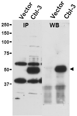
- Immunoprecipitation and western blot using Affinity Purified anti-Cbl-c antibody shows detection of a pre-dominant band at ~52 kDa corresponding to Cbl-c (arrowhead) in transfected cell lysates (left panel). Lysates are from Hek 293T cells transfected with empty vector or with Cbl-c. The predicted size of Cbl-c is 52 kDa. Size markers in kDa are shown to the left of the panel. The right panel shows western blotting after first immunoprecipitating with Rabbit anti-Cbl-c followed by immunoblotting using a Goat anti-Cbl-c antibody.
Supportive validation
- Submitted by
- Acris Antibodies GmbH (provider)
- Main image

- Experimental details
- Immunohistochemistry. Affinity Purified anti-Cbl-c antibody shows strong nuclear and cytoplasmic staining of cells in tubuli in human kidney tissue. Tissue was formalin-fixed and paraffin embedded. Brown color indicates presence of protein, blue color shows cell nuclei.

- Submitted by
- Acris Antibodies GmbH (provider)
- Main image

- Experimental details
- Immunohistochemistry. Affinity purified anti-Cbl-c antibody was used at 5 µg/ml to detect signal in a variety of tissues including multi-human, multi-brain and multi-cancer slides. This image shows moderate intracellular positive staining of human pancreatic acinar epithelium at 40X. Tissue was formalin-fixed and paraffin embedded. The image shows localization of the antibody as the precipitated red signal, with a hematoxylin purple nuclear counterstain.
