Explore
 Validate
 Learn
Learn Western blot
 Immunocytochemistry
Antibody data
- Antibody Data
- Antigen structure
- References [6]
- Comments [0]
- Validations
- Immunocytochemistry [1]
- Immunohistochemistry [1]
Submit
Validation data
Reference
Comment
Report error
- Product number
- AF1384 - Provider product page

- Provider
- R&D Systems
- Product name
- Human sFRP-1 Antibody
- Antibody type
- Polyclonal
- Description
- Antigen Affinity-purified. Detects human sFRP-1 in direct ELISAs and Western blots. In direct ELISAs, less than 2% cross-reactivity with recombinant human (rh) sFRP-2, and rhsFRP-5 is observed.
- Reactivity
- Human
- Host
- Goat
- Conjugate
- Unconjugated
- Antigen sequence
AAB70793- Isotype
- IgG
- Vial size
- 100 ug
- Concentration
- LYOPH
- Storage
- Use a manual defrost freezer and avoid repeated freeze-thaw cycles. 12 months from date of receipt, -20 to -70 °C as supplied. 1 month, 2 to 8 °C under sterile conditions after reconstitution. 6 months, -20 to -70 °C under sterile conditions after reconstitution.
Submitted references Forced expression of Wnt antagonists sFRP1 and WIF1 sensitizes chronic myeloid leukemia cells to tyrosine kinase inhibitors.
Low expression of RBMS3 and SFRP1 are associated with poor prognosis in patients with gastric cancer.
Blockade of Wnt signaling inhibits angiogenesis and tumor growth in hepatocellular carcinoma.
Wnt signaling mediates experience-related regulation of synapse numbers and mossy fiber connectivities in the adult hippocampus.
Wnt signaling antagonists are potential prognostic biomarkers for the progression of radiographic hip osteoarthritis in elderly Caucasian women.
Genomics identifies medulloblastoma subgroups that are enriched for specific genetic alterations.
Pehlivan M, Caliskan C, Yuce Z, Sercan HO
Tumour biology : the journal of the International Society for Oncodevelopmental Biology and Medicine 2017 May;39(5):1010428317701654
Tumour biology : the journal of the International Society for Oncodevelopmental Biology and Medicine 2017 May;39(5):1010428317701654
Low expression of RBMS3 and SFRP1 are associated with poor prognosis in patients with gastric cancer.
Zhang T, Wu Y, Fang Z, Yan Q, Zhang S, Sun R, Khaliq J, Li Y
American journal of cancer research 2016;6(11):2679-2689
American journal of cancer research 2016;6(11):2679-2689
Blockade of Wnt signaling inhibits angiogenesis and tumor growth in hepatocellular carcinoma.
Hu J, Dong A, Fernandez-Ruiz V, Shan J, Kawa M, Martínez-Ansó E, Prieto J, Qian C
Cancer research 2009 Sep 1;69(17):6951-9
Cancer research 2009 Sep 1;69(17):6951-9
Wnt signaling mediates experience-related regulation of synapse numbers and mossy fiber connectivities in the adult hippocampus.
Gogolla N, Galimberti I, Deguchi Y, Caroni P
Neuron 2009 May 28;62(4):510-25
Neuron 2009 May 28;62(4):510-25
Wnt signaling antagonists are potential prognostic biomarkers for the progression of radiographic hip osteoarthritis in elderly Caucasian women.
Lane NE, Nevitt MC, Lui LY, de Leon P, Corr M, Study of Osteoporotic Fractures Research Group.
Arthritis and rheumatism 2007 Oct;56(10):3319-25
Arthritis and rheumatism 2007 Oct;56(10):3319-25
Genomics identifies medulloblastoma subgroups that are enriched for specific genetic alterations.
Thompson MC, Fuller C, Hogg TL, Dalton J, Finkelstein D, Lau CC, Chintagumpala M, Adesina A, Ashley DM, Kellie SJ, Taylor MD, Curran T, Gajjar A, Gilbertson RJ
Journal of clinical oncology : official journal of the American Society of Clinical Oncology 2006 Apr 20;24(12):1924-31
Journal of clinical oncology : official journal of the American Society of Clinical Oncology 2006 Apr 20;24(12):1924-31
No comments: Submit comment
Supportive validation
- Submitted by
- R&D Systems (provider)
- Main image

- Experimental details
- sFRP-1 in MBA-MB-468 Human Cell Line. sFRP-1 was detected in immersion fixed MBA-MB-468 human breast cancer cell line using Goat Anti-Human sFRP-1 Antigen Affinity-purified Polyclonal Antibody (Catalog # AF1384) at 15 µg/mL for 3 hours at room temperature. Cells were stained using the NorthernLights™ 557-conjugated Anti-Goat IgG Secondary Antibody (red; Catalog # NL001) and counterstained with DAPI (blue). Specific staining was localized to cytoplasm. View our protocol for Fluorescent ICC Staining of Cells on Coverslips.

Supportive validation
- Submitted by
- R&D Systems (provider)
- Main image

- Experimental details
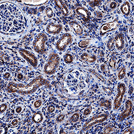
- sFRP-1 in Human Kidney. sFRP-1 was detected in immersion fixed paraffin-embedded sections of human kidney using Goat Anti-Human sFRP-1 Antigen Affinity-purified Polyclonal Antibody (Catalog # AF1384) at 1 µg/mL overnight at 4 °C. Tissue was stained using the Anti-Goat HRP-DAB Cell & Tissue Staining Kit (brown; Catalog # CTS008) and counterstained with hematoxylin (blue). Specific staining was localized to epithelial cell cytoplasm in convoluted tubules. View our protocol for Chromogenic IHC Staining of Paraffin-embedded Tissue Sections.