Explore
 Validate
 Learn
Learn Western blot
 Immunoprecipitation
Antibody data
- Antibody Data
- Antigen structure
- References [5]
- Comments [0]
- Validations
- Western blot [3]
- Immunohistochemistry [1]
- Flow cytometry [1]
Submit
Validation data
Reference
Comment
Report error
- Product number
- NB100-456 - Provider product page

- Provider
- Novus Biologicals
- Proper citation
- Novus Cat#NB100-456, RRID:AB_10001776
- Product name
- Rabbit Polyclonal PCNA Antibody
- Antibody type
- Polyclonal
- Description
- Immunogen affinity purified.
- Reactivity
- Human, Mouse
- Host
- Rabbit
- Isotype
- IgG
- Vial size
- 100 ul
- Concentration
- 1.0 mg/ml
- Storage
- Store at 4C. Do not freeze.
Submitted references BMP4 induces asymmetric cell division in human glioma stem-like cells.
Epidermal growth factor receptor protects proliferating cell nuclear antigen from cullin 4A protein-mediated proteolysis.
The E3 ubiquitin ligase RAD18 regulates ubiquitylation and chromatin loading of FANCD2 and FANCI.
Processing of lagging-strand intermediates in vitro by herpes simplex virus type 1 DNA polymerase.
HGF mediates cell proliferation of human mesothelioma cells through a PI3K/MEK5/Fra-1 pathway.
Koguchi M, Nakahara Y, Ito H, Wakamiya T, Yoshioka F, Ogata A, Inoue K, Masuoka J, Izumi H, Abe T
Oncology letters 2020 Feb;19(2):1247-1254
Oncology letters 2020 Feb;19(2):1247-1254
Epidermal growth factor receptor protects proliferating cell nuclear antigen from cullin 4A protein-mediated proteolysis.
Lo YH, Ho PC, Wang SC
The Journal of biological chemistry 2012 Aug 3;287(32):27148-57
The Journal of biological chemistry 2012 Aug 3;287(32):27148-57
The E3 ubiquitin ligase RAD18 regulates ubiquitylation and chromatin loading of FANCD2 and FANCI.
Williams SA, Longerich S, Sung P, Vaziri C, Kupfer GM
Blood 2011 May 12;117(19):5078-87
Blood 2011 May 12;117(19):5078-87
Processing of lagging-strand intermediates in vitro by herpes simplex virus type 1 DNA polymerase.
Zhu Y, Wu Z, Cardoso MC, Parris DS
Journal of virology 2010 Aug;84(15):7459-72
Journal of virology 2010 Aug;84(15):7459-72
HGF mediates cell proliferation of human mesothelioma cells through a PI3K/MEK5/Fra-1 pathway.
Ramos-Nino ME, Blumen SR, Sabo-Attwood T, Pass H, Carbone M, Testa JR, Altomare DA, Mossman BT
American journal of respiratory cell and molecular biology 2008 Feb;38(2):209-17
American journal of respiratory cell and molecular biology 2008 Feb;38(2):209-17
No comments: Submit comment
Supportive validation
- Submitted by
- Novus Biologicals (provider)
- Main image
- Experimental details
- Western Blot: PCNA Antibody [NB100-456] - A431 whole cell lysates
- Submitted by
- Novus Biologicals (provider)
- Main image
- Experimental details
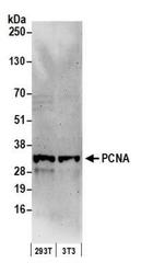
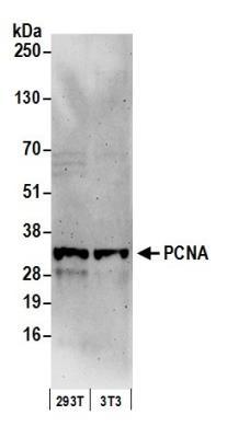
- Western Blot: PCNA Antibody [NB100-456] - Detection of Human and Mouse PCNA by Western Blot. Samples: Whole cell lysate (50 ug) from 293T and mouse NIH3T3 cells prepared using NETN lysis buffer. Antibody: Affinity purified rabbit anti-PCNA antibody NB100-456 used for WB at 0.1 ug/ml. Detection: Chemiluminescence with an exposure time of 3 minutes.
- Submitted by
- Novus Biologicals (provider)
- Main image

- Experimental details
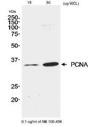
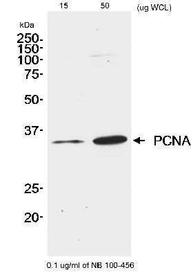
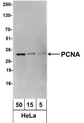
- Western Blot: PCNA Antibody [NB100-456] - Analysis using the Biotin conjugate of NB100-456. Detection ofHeLa (50, 15, and 5 ug). Antibody used at 1.0 ug/ml. Detection: Streptavidin-HRP and chemiluminescence with an exposure time of 3 seconds.
Supportive validation
- Submitted by
- Novus Biologicals (provider)
- Main image
- Experimental details
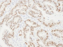
- Immunohistochemistry: PCNA Antibody [NB100-456] - Sample: FFPE section of human stomach carcinoma. Antibody: Affinity purified rabbit anti- PCNA used at a dilution of 1:10,000 (0.1ug/ml). Detection: DAB

Supportive validation
- Submitted by
- Novus Biologicals (provider)
- Main image

- Experimental details
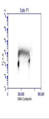
- Flow Cytometry: PCNA Antibody [NB100-456] - Detection of PCNA Versus DNA Content. Asynchronous Jurkat cells were fixed and permeabilized in a sequential treatment of FACS buffer (PBS, 0.5% triton-X-100, 0.5mM EDTA, 1% BSA) and 100% methanol. 1 X10⁶ cells were stained with 0.03 ug anti-PCNA . Secondary detection was performed with FITC conjugated Goat F(ab')2 anti-rabbit antibody], and DNA stained with PI.