Explore
 Validate
 Learn
Learn Western blot
 ELISA
ELISAAntibody data
- Antibody Data
- Antigen structure
- References [0]
- Comments [0]
- Validations
- Western blot [1]
- Immunohistochemistry [1]
Submit
Validation data
Reference
Comment
Report error
- Product number
- AP09256PU-N - Provider product page

- Provider
- Acris Antibodies GmbH
- Proper citation
- Acris Antibodies GmbH Cat#AP09256PU-N, RRID:AB_2035441
- Product name
- anti E2F1 pSer364
- Antibody type
- Polyclonal
- Antigen
- Synthetic peptide corresponding to amino acids 360-369 of Human E2F-1
- Reactivity
- Human
- Host
- Rabbit
- Isotype
- IgG
- Vial size
- 0.1 mg
- Concentration
- 0.80 mg/ml (by UV absorbance at 280 nm)
No comments: Submit comment
Supportive validation
- Submitted by
- Acris Antibodies GmbH (provider)
- Main image

- Experimental details
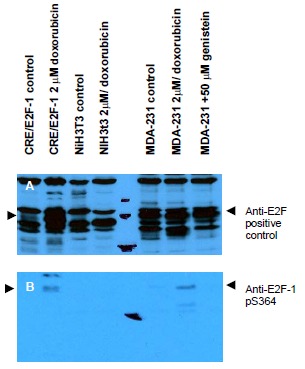
- Western blot using anti-E2F-1 pS364 antibody shows detection of a band at ~47 kDa corresponding to phosphorylated E2F-1 in induced cell lysates. Panel A shows reactivity using a control antibody reactive to all forms of E2F (arrowheads). Panel B shows specific reactivity against phosphorylated E2F-1 (arrowheads) using our anti-E2F1 pS364 antibody. Lysates are as follows: CRE/E2F-1 are CRE cells derived from mouse NIH3T3 line transfected with human E2F-1, NIH-3T3 used as a negative control, and MDA-MB-231 cells are a human breast cancer line. As indicated each lysatewas prepared from untreated cells and cells treated with 2 μM Doxorubicin used as a DNA damaging agent. In addition the MDA-MB-231 cells were also treated with genistein, a mild Ddamaging agent. The figure shows the same membrane first probed with the anti-E2F-1 pS364 antibody used at a 1:250 dilution, then stripped and re-probed with the pan E2F antibodyused as a positive control. The positive control antibody clearlyshows an E2F-1 band in all human cell lines, but not mouse cells. Treatment with doxorubicin increases the expression oE2F-1 as shown in Panel A. After film development, images were overlapped to confirm that specific anti-E2F-1 pS364 staining shown treated human cells in Panel B specifically aligns with E2F-1 staining shown in Panel A. Blots can be processed with HRP conjugated Gt-a-Rabbit IgG for 45 min at room temperature for ECL detection. Personal Communication, XiaoHe Yang, Univ. Oklahoma.
Supportive validation
- Submitted by
- Acris Antibodies GmbH (provider)
- Main image

- Experimental details
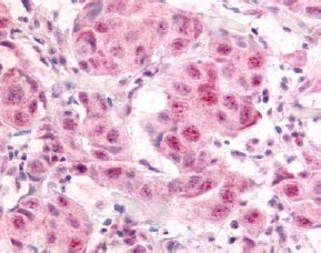
- Immunohistochemistry. Anti-E2F-1 pS364 antibody was used at a 10 μg/ml to detect nuclear and occasion-ally cytoplasmic signal in a variety of tissues in-cluding multi-human, multi-brain and multi-cancer slides. Within the multi-tumor block, the antibody showed variable levels of nuclear staining between individual tumors, with some tumors showing strong staining. This image shows E2F-1 pS364 staining of human breast carcinoma. Tissue was formalin-fixed and paraffin embedded. Personal Communication, Tina Roush, LifeSpanBiosciences, Seattle, WA.