Explore
 Validate
 Learn
Learn Immunocytochemistry
Antibody data
- Antibody Data
- Antigen structure
- References [0]
- Comments [0]
- Validations
- Immunocytochemistry [2]
- Immunohistochemistry [6]
Submit
Validation data
Reference
Comment
Report error
- Product number
- AMAb91297 - Provider product page

- Provider
- Atlas Antibodies
- Proper citation
- Atlas Antibodies Cat#AMAb91297, RRID:AB_2665884
- Product name
- Anti-SOX10
- Antibody type
- Monoclonal
- Reactivity
- Human, Mouse
- Host
- Mouse
- Conjugate
- Unconjugated
- Antigen sequence
PHYTDQPSTSQIAYTSLSLPHYGSAFPSISRPQFD
YSDHQPSGPYYGHSG- Isotype
- IgG
- Antibody clone number
- CL4455
- Vial size
- 100 µl
- Storage
- Store at +4°C for short term storage. Long time storage is recommended at -20°C.
No comments: Submit comment
Supportive validation
- Submitted by
- Atlas Antibodies (provider)
- Main image
- Experimental details
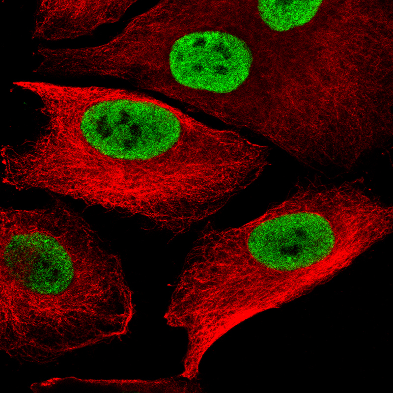
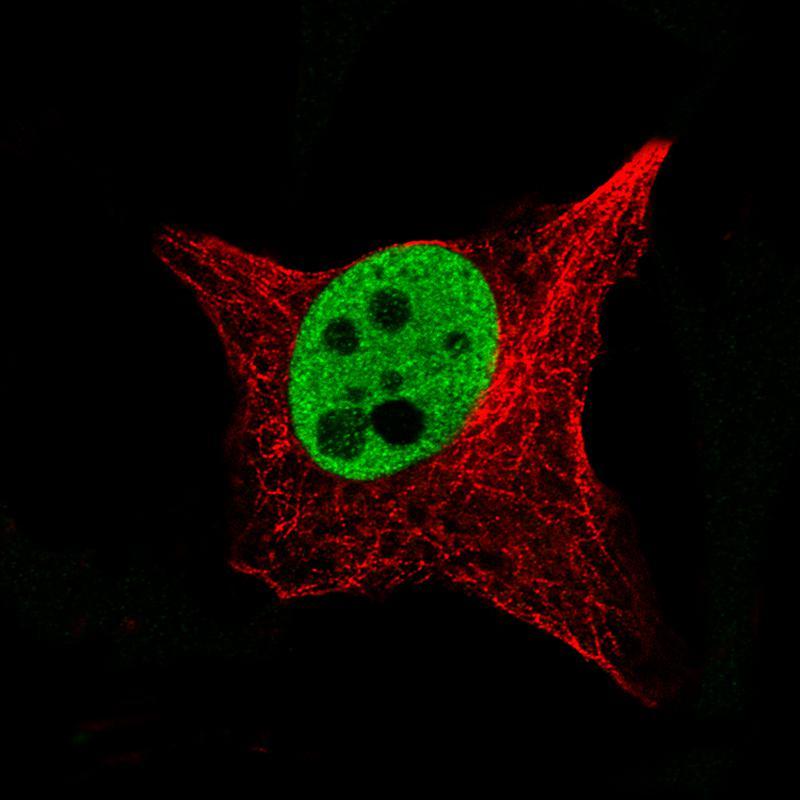
- Immunofluorescence staining of WM-115 cells using the anti-SOX10 monoclonal antibody, showing specific staining in the nucleoplasm in green. Microtubule- and nuclear probes are visualized in red and blue, respectively (where available).
- Submitted by
- Atlas Antibodies (provider)
- Main image
- Experimental details
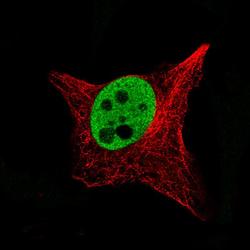
- Immunofluorescence staining of SK-MEL-30 cells using the Anti-SOX10 monoclonal antibody, showing specific staining in the nucleoplasm in green. Microtubule- and nuclear probes are visualized in red and blue, respectively (where available).
- Sample type
- HUMAN
Supportive validation
- Submitted by
- Atlas Antibodies (provider)
- Main image
- Experimental details
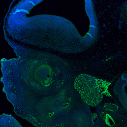
- Immunofluorescence staining of mouse embryo E11 shows nuclear immunoreactivity in the developing trigeminal ganglion and ear placode.

- Submitted by
- Atlas Antibodies (provider)
- Main image
- Experimental details
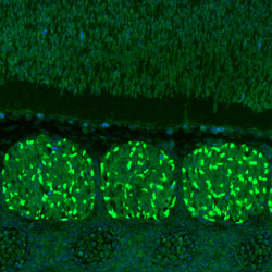
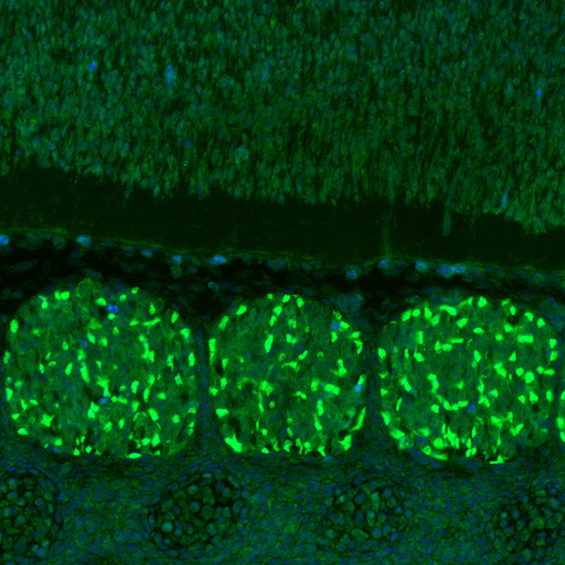
- Immunofluorescence staining of mouse embryo E11 shows nuclear immunoreactivity in the developing dorsal root ganglia.
- Submitted by
- Atlas Antibodies (provider)
- Main image
- Experimental details
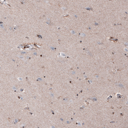
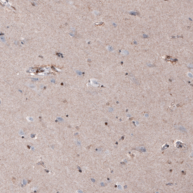
- Immunohistochemical staining of human cerebral cortex shows strong nuclear immunoreactivity in a subset of glial cells.
- Submitted by
- Atlas Antibodies (provider)
- Main image
- Experimental details
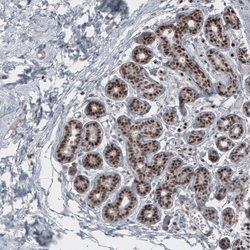
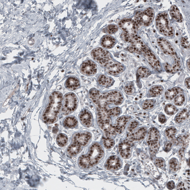
- Immunohistochemical staining of human breast shows strong nuclear positivity in glandular cells.
- Submitted by
- Atlas Antibodies (provider)
- Main image
- Experimental details
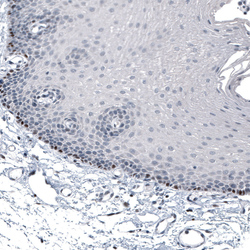
- Immunohistochemical staining of human cervix shows nuclear immunoreactivity in a subset of basal cells in squamous epithelium.

- Submitted by
- Atlas Antibodies (provider)
- Main image
- Experimental details
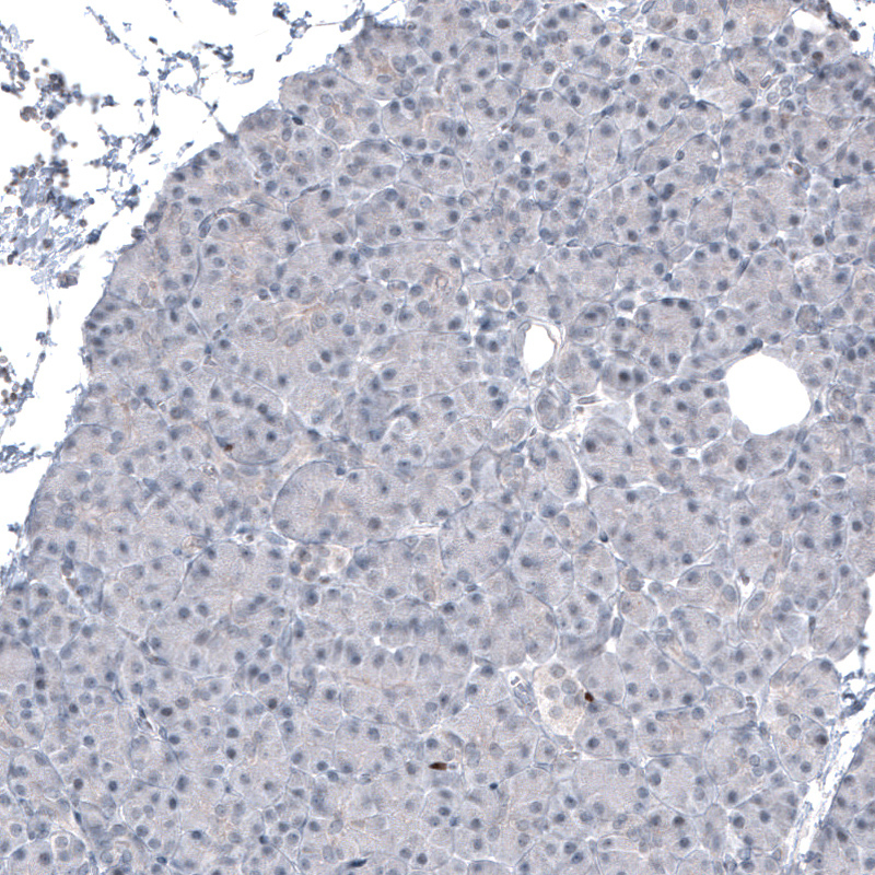
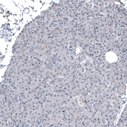
- Immunohistochemical staining of human pancreas shows absence of positivity as expected (negative control).