Explore
 Validate
 Learn
Learn Western blot
 Flow cytometry
Antibody data
- Antibody Data
- Antigen structure
- References [0]
- Comments [0]
- Validations
- Flow cytometry [1]
Submit
Validation data
Reference
Comment
Report error
- Product number
- NBP2-59621AF488 - Provider product page

- Provider
- Novus Biologicals
- Product name
- Mouse Monoclonal SOX10 Antibody
- Antibody type
- Monoclonal
- Description
- Protein A or G purified. The specificity of this monoclonal antibody to human, mouse SOX10 was validated by HuProtTM Array, containing more than 19,000, full-length human proteins. Recognizes a protein of ~55kDa, identified as SOX10. This MAb is highly specific and does not cross-react with other members of the SOX-family. SOX genes comprise a family of genes that are related to the mammalian sex-determining gene SRY. These genes similarly contain sequences that encode for the HMG -box domain, which is responsible for the sequence-specific DNA-binding activity. SOX-10 is a sensitive marker of melanoma, including conventional, spindled, and desmoplastic subtypes. It is expressed by metastatic melanomas and nodal capsular nevus in sentinel lymph nodes, but not by other lymph node components such as dendritic cells, which usually express S100 protein. Commonly used melanoma markers, such as anti-HMB-45 and anti-Melan-A, are poorly expressed in desmoplastic melanomas while SOX-10 is moderately to strongly expressed in desmoplastic melanomas. SOX-10 is considered as a very reliable marker for recognizing residual desmoplastic melanomas. In normal tissues, it is expressed in Schwann cells, melanocytes, and myoepithelial cells of salivary, bronchial and mammary glands. SOX-10 expression is also observed in mast cells
- Reactivity
- Human, Mouse
- Host
- Mouse
- Conjugate
- Green dye
- Isotype
- IgG
- Vial size
- 100 ul
- Storage
- Store at 4C in the dark.
No comments: Submit comment
Supportive validation
- Submitted by
- Novus Biologicals (provider)
- Main image

- Experimental details
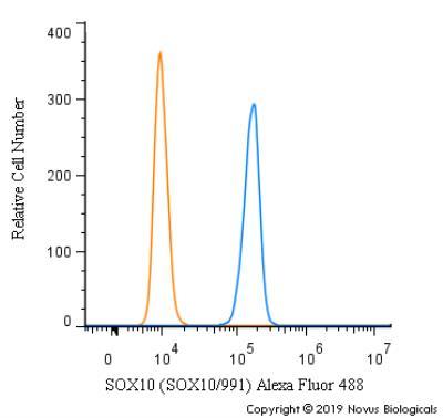
- Flow Cytometry: SOX10 Antibody (SOX10/991) [Alexa Fluor® 488] [NBP2-59621AF488] - An intracellular stain was performed on SK-MEL-28 cells with SOX10 [SOX10/991] Antibody NBP2-59621AF488 (blue) and a matched isotype control (orange). Cells were fixed with 4% PFA and then permeabilized with 0.1% saponin. Cells were incubated in an antibody dilution of 5 ug/mL for 30 minutes at room temperature. Both antibodies were conjugated to Alexa Fluor 488.